

doi: 10.5009/gnl.2010.4.1.117.
Epub 2010 Mar 25.
Ileal Mucosa-Associated Lymphoid Tissue (MALT) Lymphoma with a Large-Cell Component That Regressed Spontaneously
Affiliations
- PMID: 20479924
- PMCID: PMC2871597
- DOI: 10.5009/gnl.2010.4.1.117
Item in Clipboard
Ileal Mucosa-Associated Lymphoid Tissue (MALT) Lymphoma with a Large-Cell Component That Regressed Spontaneously
Gut Liver.
2010 Mar.
Abstract
Reported herein is a case of mucosa-associated lymphoid tissue (MALT) lymphoma of the terminal ileum with a large-cell component, which regressed spontaneously. To the best of our knowledge, only five cases of spontaneously regressing MALT lymphoma have been reported in the English-language literature, and all of these cases were low-grade lymphomas. Spontaneous regression of a MALT lymphoma with a high-grade component is very rare. The present case suggests that MALT lymphoma cells have a reversible nature, even in the presence of a high-grade component.
Keywords: Ileum, Low grade; Large cell component; Mucosa-associated lymphoid tissue lymphoma; Regression.
Figures
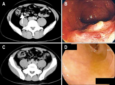
Radiological and endoscopic findings of an ileal lesion in one patient. (A) February 3, 2007: computed tomography (CT) finding of slightly enlarged ileocecal lymph nodes (arrow). (B) February 16, 2007: colonoscopic examination disclosed multiple protruding lesions with erosive tops and covered by edematous mucosa in the terminal ileum. (C) March 23, 2007: CT findings of no detectable ileocecal lymph nodes. (D) April 5, 2007: no lesions could be found in the terminal ileum on colonoscopic examination.
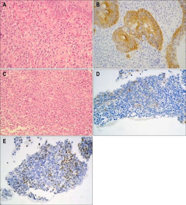
Pathological findings of ileal lesions. (A) Dense, homogenous, diffuse mononuclear cell infiltration was demonstrated in biopsy specimens of the terminal ileal mucosa (H&E stain, original magnification, ×100). (B) Lymphoepithelial lesions were clearly detected by immunohistochemistry against cytokeratin AE1/AE3 (DAKO; original magnification, ×400). (C) Diffuse large-B-cell lymphoma-like cells with swollen, light-colored nuclei and a high nuclear-cytoplasmic ratio in a biopsy specimen of the terminal ileum. (D) Immunohistochemistry against CD10 (original magnification, ×400). (E) Immunohistochemistry against BCL2 (original magnification, ×400).
Similar articles
-
Clinicopathological features of gastric mucosa-associated lymphoid tissue lymphoma: a comparison with diffuse large B-cell lymphoma without a mucosa-associated lymphoid tissue lymphoma component.J Gastroenterol Hepatol. 2001 Jul;16(7):734-9. doi: 10.1046/j.1440-1746.2001.02519.x. J Gastroenterol Hepatol. 2001. PMID: 11446880
-
Evaluation of CD23 expression in paraffin-embedded gastric lymphomas of mucosa-associated lymphoid tissue.Mod Pathol. 1998 Oct;11(10):967-70. Mod Pathol. 1998. PMID: 9796724
-
Mucosal change of the stomach with low-grade mucosa-associated lymphoid tissue lymphoma after eradication of Helicobacter pylori: follow-up study of 48 cases.J Med Invest. 2000 Feb;47(1-2):36-46. J Med Invest. 2000. PMID: 10740978
-
Recent developments in our understanding of gastric lymphomas.Am J Surg Pathol. 1996;20 Suppl 1:S1-7. doi: 10.1097/00000478-199600001-00002. Am J Surg Pathol. 1996. PMID: 8694145 Review.
-
Mucosa-associated lymphoid tissue (MALT) variant of primary rectal lymphoma: a review of the English literature.Int J Colorectal Dis. 2017 Mar;32(3):295-304. doi: 10.1007/s00384-016-2734-z. Epub 2016 Dec 19. Int J Colorectal Dis. 2017. PMID: 27995323 Review.
Cited by
-
Mucosa-Associated Lymphoid Tissue (MALT) Lymphoma in the Gastrointestinal Tract in the Modern Era.Cancers (Basel). 2022 Jan 17;14(2):446. doi: 10.3390/cancers14020446. Cancers (Basel). 2022. PMID: 35053607 Free PMC article. Review.
-
Role of F-FDG PET Scans in Patients with Helicobacter pylori-Infected Gastric Low-Grade MALT Lymphoma.Gut Liver. 2011 Sep;5(3):308-14. doi: 10.5009/gnl.2011.5.3.308. Epub 2011 Aug 18. Gut Liver. 2011. PMID: 21927659 Free PMC article.
-
A journey into insidious world of MALT lymphoma of the ileum: from the beginning to the end.J Gastrointest Oncol. 2014 Dec;5(6):E125-7. doi: 10.3978/j.issn.2078-6891.2014.063. J Gastrointest Oncol. 2014. PMID: 25436136 Free PMC article.
-
Spontaneous regression of primary extranodal marginal zone lymphoma of mucosa-associated lymphoid tissue (MALT lymphoma) colliding with invasive ductal carcinoma of the breast: a case report.Int J Clin Exp Pathol. 2014 Sep 15;7(10):7020-7. eCollection 2014. Int J Clin Exp Pathol. 2014. PMID: 25400790 Free PMC article.
-
Mucosa-associated lymphoid tissue lymphoma in the terminal ileum: A case report.World J Gastrointest Endosc. 2022 Mar 16;14(3):176-182. doi: 10.4253/wjge.v14.i3.176. World J Gastrointest Endosc. 2022. PMID: 35432742 Free PMC article.
References
-
- Isaacson P, Wright DH. Malignant lymphoma of mucosa-associated lymphoid tissue: a distinctive type of B-cell lymphoma. Cancer. 1983;52:1410–1416. - PubMed
-
- Isaacson PG. The MALT lymphoma concept updated. Ann Oncol. 1995;6:319–320. - PubMed
-
- Radaszkiewicz T, Dragosics B, Bauer P. Gastrointestinal malignant lymphomas of the mucosa-associated lymphoid tissue: factors relevant to prognosis. Gastroenterology. 1992;102:1628–1638. - PubMed
-
- Cogliatti SB, Schmid U, Schumacher U, et al. Primary B-cell gastric lymphoma: a clinicopathological study of 145 patients. Gastroenterology. 1991;101:1159–1170. - PubMed
-
- de Jong D, Boot H, van Heerde P, Hart GA, Taal BG. Histological grading in gastric lymphoma: pretreatment criteria and clinical relevance. Gastroenterology. 1997;112:1466–1474. - PubMed
LinkOut - more resources
Full Text Sources
