

Inflammatory myoglandular polyps causing hematochezia
- PMID: 20479930
- PMCID: PMC2871605
- DOI: 10.5009/gnl.2010.4.1.146
Inflammatory myoglandular polyps causing hematochezia
Abstract
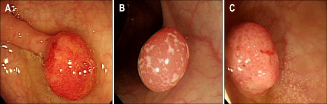
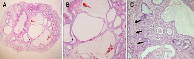
We report herein three cases of inflammatory myoglandular polyp (IMGP) presenting as hematochezia. The polyps had pedunculated, red, and smooth features, and were 12, 12, and 15 mm in diameter and located in the sigmoid colon, transverse colon, and rectum, respectively. Endoscopic polypectomies were performed. Histologic examination of the recovered specimens revealed inflammatory granulation in the lamina propria mucosa, proliferation of smooth muscle, and hyperplastic glands with cystic dilatation. The three colon polyps were finally diagnosed both clinically and histologically as IMGP. Endoscopists should bear in mind that a polyp featuring endoscopic findings of pedunculation or semipedunculation; a red, smooth, spherical, and hyperemic surface; and patchy mucosa exudation and erosion is likely to be an IMGP.
Keywords: Endoscopic polypectomy; Gastrointestinal hemorrhage; Inflammatory myoglandular polyp.
Figures
References
-
- Nakamura S, Kino I, Akagi T. Inflammatory myoglandular polyps of the colon and rectum: a clinicopathological study of 32 pedunculated polyps, distinct from other types of polyps. Am J Surg Pathol. 1992;16:772–779. - PubMed
-
- Bhathal PS, Chetty R, Slavin JL. Myoglandular polyps. Am J Surg Pathol. 1993;17:852–853. - PubMed
-
- Griffiths AP, Hopkinson JM, Dixon MF. Inflammatory myoglandular polyp causing ileo-ileal intussusception. Histopathology. 1993;23:596–598. - PubMed
-
- Gomez Navarro E, del Rio Martin JV, Sarasa Corral JL, Melero Calleja E. Myoglandular inflammatory polyp located in the distal end of the rectum. Rev Esp Enferm Dig. 1994;85:45–46. - PubMed
-
- Nagata S, Sumioka M, Sato O, et al. Five cases of inflammatory myoglandular polyp. Nippon Shokakibyo Gakkai Zasshi. 1998;95:145–150. - PubMed
LinkOut - more resources
Full Text Sources
