. 2010 May 10;5(5):e10566.
doi: 10.1371/journal.pone.0010566.

Serum anti-BPAG1 auto-antibody is a novel marker for human melanoma

Affiliations

Serum anti-BPAG1 auto-antibody is a novel marker for human melanoma

Takashi Shimbo et al. PLoS One. .

Abstract

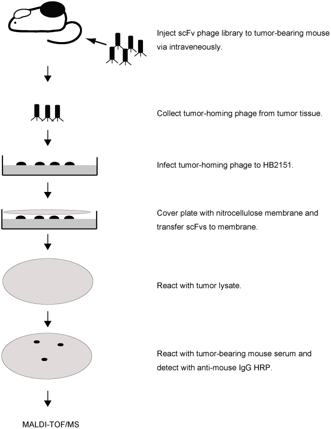
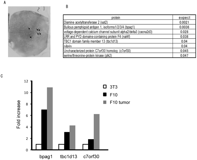
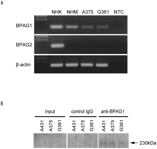
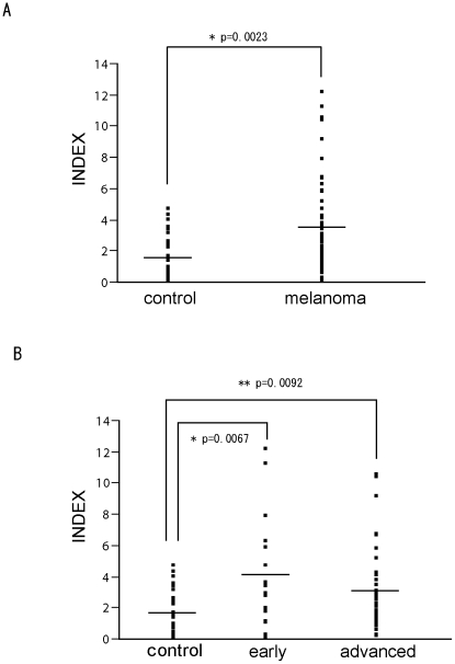
Malignant melanoma is one of the most aggressive types of tumor. Because malignant melanoma is difficult to treat once it has metastasized, early detection and treatment are essential. The search for reliable biomarkers of early-stage melanoma, therefore, has received much attention. By using a novel method of screening tumor antigens and their auto-antibodies, we identified bullous pemphigoid antigen 1 (BPAG1) as a melanoma antigen recognized by its auto-antibody. BPAG1 is an auto-antigen in the skin disease bullous pemphigoid (BP) and anti-BPAG1 auto-antibodies are detectable in sera from BP patients and are used for BP diagnosis. However, BPAG1 has been viewed as predominantly a keratinocyte-associated protein and a relationship between BPAG1 expression and melanoma has not been previously reported. In the present study, we show that bpag1 is expressed in the mouse F10 melanoma cell line in vitro and F10 melanoma tumors in vivo and that BPAG1 is expressed in human melanoma cell lines (A375 and G361) and normal human melanocytes. Moreover, the levels of anti-BPAG1 auto-antibodies in the sera of melanoma patients were significantly higher than in the sera of healthy volunteers (p<0.01). Furthermore, anti-BPAG1 auto-antibodies were detected in melanoma patients at both early and advanced stages of disease. Here, we report anti-BPAG1 auto-antibodies as a promising marker for the diagnosis of melanoma, and we discuss the significance of the detection of such auto-antibodies in cancer biology and patients.


Conflict of interest statement

Competing Interests: The authors have declared that no competing interests exist.

Figures

Figure 1
Figure 1. Overview of the rapid method for isolating auto-antibody against tumor-associated antigen (TAA) using a scFv library.
The tumor-homing scFv-presenting phages were collected from tumors that were injected with a scFv library. The collected phages were infected to HB2151 for scFv secretion. The secreted scFvs from HB2151 were transferred to nitrocellulose membranes by colony lift. The membranes were incubated with tumor lysate followed by serum from a tumor-bearing mouse. The scFv-tumor protein complex was detected by auto-antibodies. The complex was digested into peptide by trypsin and analyzed using MALDI-TOF mass spectrometry for identification.
Figure 2
Figure 2. Identification of bpag1 as a tumor antigen recognized by auto-antibodies.
(A) An example of the screening output. ScFv-tumor antigen complex was detected with auto-antibodies in tumor-bearing mouse serum. (B) Eight candidates were identified by MALDI-TOF mass spectrometry with statistical significance (p<0.05); expect  =  expectation value. (C) Comparison of bpag1, tbc1d13 and c7orf30 expression in NIH-3T3 cells (white bar), F10 melanoma cells (black bar) and F10 melanoma tumors (grey bar) by SYBR Green real-time PCR.
Figure 3
Figure 3. Expression of BPAG1 in normal human melanocytes and human melanoma cell lines.
(A) The expression of BPAG1 and BPAG2 mRNA was quantified by RT-PCR in normal human melanocytes (NHM) and human melanoma cell lines A375 and G361. Normal human keratinocyte (NHK) mRNA was used as a positive control. β-actin was amplified as a loading control for cDNA. NTC; no template control. (B) The expression of BPAG1 protein was detected by IP-western blotting in human melanoma cell lines A375 and G361. A431 was used as positive control for BPAG1. The arrow indicates BPAG1.
Figure 4
Figure 4. Quantification of anti-BPAG1 auto-antibodies in melanoma patients.
(A) The levels of anti-BPAG1 auto-antibodies in sera collected from healthy volunteers and melanoma patients were quantified using a MESACUP BP230 ELISA Kit. The INDEX values were plotted and the average INDEX values as shown (±S.E.M.) for control subjects (1.64±0.27) and melanoma patients (3.47±0.40). (B) The melanoma patients were classified using the American Joint Committee on Cancer (AJCC) 2002 staging criteria. In situ, stage I or stage II patients were categorized as “early”, while stage III or stage IV patients were categorized as “advanced”. The average INDEX values (±S.E.M.) of early and advanced melanoma patients were 4.14±0.83 and 3.15±0.43, respectively; the bars indicate the average INDEX value.

Similar articles

Cited by

References

    1. Jemal A, Siegel R, Ward E, Hao Y, Xu J, et al. Cancer statistics, 2008. CA Cancer J Clin. 2008;58:71–96. - PubMed
    1. Bosserhoff AK. Novel biomarkers in malignant melanoma. Clin Chim Acta. 2006;367:28–35. - PubMed
    1. Gould Rothberg BE, Bracken MB, Rimm DL. Tissue biomarkers for prognosis in cutaneous melanoma: a systematic review and meta-analysis. J Natl Cancer Inst. 2009;101:452–474. - PMC - PubMed
    1. Ohsie SJ, Sarantopoulos GP, Cochran AJ, Binder SW. Immunohistochemical characteristics of melanoma. J Cutan Pathol. 2008;35:433–444. - PubMed
    1. Brochez L, Naeyaert JM. Serological markers for melanoma. Br J Dermatol. 2000;143:256–268. - PubMed

Publication types

MeSH terms