. 2010 Sep;59(9):1367-77.
doi: 10.1007/s00262-010-0866-5. Epub 2010 May 18.

Depletion of regulatory T cells by anti-GITR mAb as a novel mechanism for cancer immunotherapy

Affiliations

Depletion of regulatory T cells by anti-GITR mAb as a novel mechanism for cancer immunotherapy

David Coe et al. Cancer Immunol Immunother. 2010 Sep.

Abstract

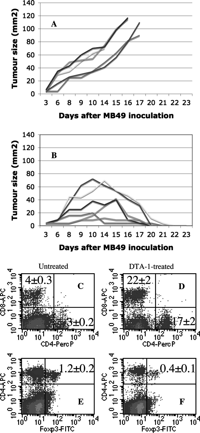
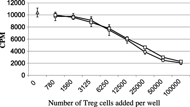
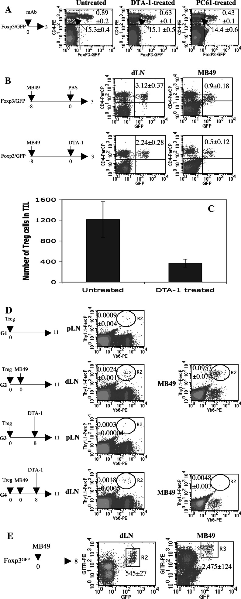
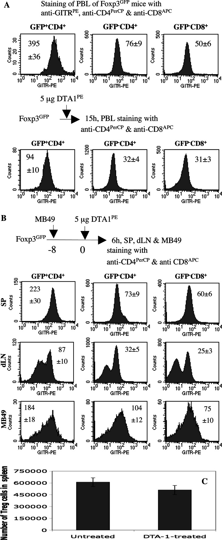
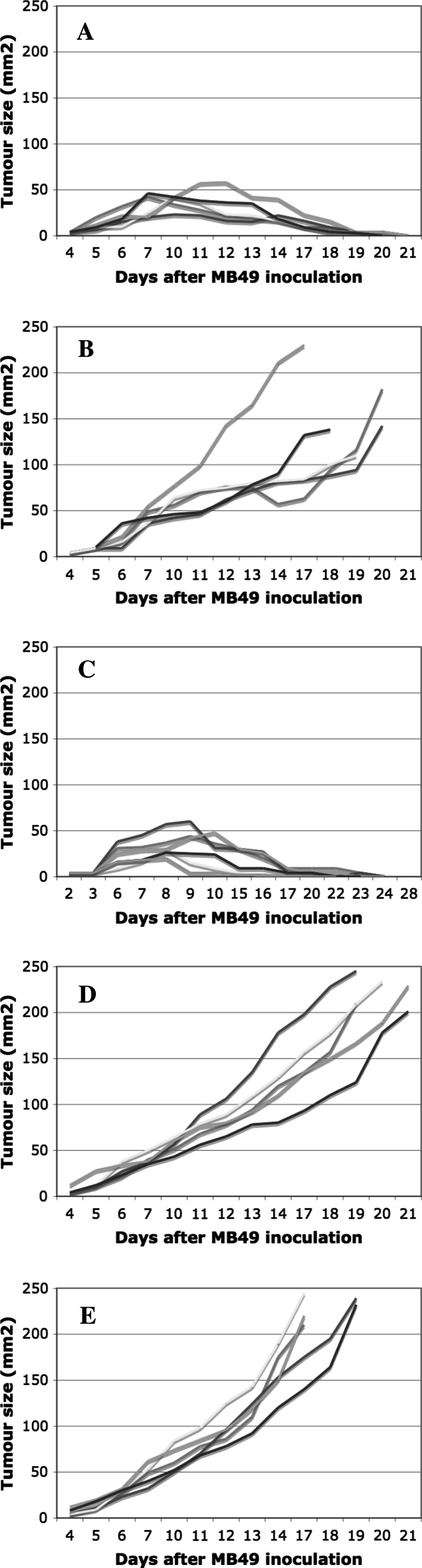
In vitro, engagement of GITR on Treg cells by the agonistic anti-GITR mAb, DTA-1, appears to abrogate their suppressive function. The consequence of in vivo engagement of GITR by DTA-1 is, however, less clear. In this study, we show that Treg cells isolated from DTA-1-treated mice were as potent as those from untreated mice in suppressing conventional CD4 T cells in vitro, indicating that in vivo GITR ligation does not disable Treg cells. Treatment of Foxp3/GFP knock-in mice with DTA-1 led to a selective reduction of circulating Treg cells, suggesting that DTA-1 is a depleting mAb which preferentially targets Treg cells. In tumour-bearing mice, DTA-1-mediated depletion of Treg cells was most marked in tumours but not in tumour-draining lymph node. These features were confirmed in an adoptive transfer model using tumour antigen-specific Treg cells. Interestingly, Treg cells detected in tumour tissues expressed much higher levels of GITR than those in tumour-draining lymph nodes, indicating that the efficiency of depletion might be correlated with the level of GITR expression. Finally, in vivo labelling of GITR in naive or tumour-bearing mice demonstrated that Treg cells constitutively expressed higher levels of GITR than conventional T cells, independent of location and activation state, consistent with the preferential in vivo depletion of Tregs by DTA-1. Thus, depletion of Treg cells represents a previously unrecognised in vivo activity of DTA-1 which has important implications for the application of anti-GITR antibodies in cancer immunotherapy.


Figures

Fig. 1
Fig. 1
DTA-1 is highly effective in treating established tumours. a, b A single injection of DTA-1 results in the regression of established MB49 tumours. A total of 11 B6 female mice were s.c. inoculated with 5 × 105 MB49 cells. 8 days later, the mice were randomly divided into two groups: one group (B, n = 6) was treated with 50 μg of DTA-1 mAb by i.p. injection and the other group was not (A, n = 5). The growth of MB49 tumours was monitored by measuring tumour size every other day. One representative of three independent experiments is shown. c–f Regression of MB49 is correlated with increased accumulation of tumour-infiltrating CD4 and CD8 T cells but decreased accumulation of Treg cells. Untreated and DTA-1-treated mice (3 mice/group) were prepared as described in A and B. At day 15, MB49 tumours isolated from untreated (C and E) and DTA-1-treated mice (D and F) were stained with anti-CD4PerCP and anti-CD8APC (C and D), or anti-CD4PerCP followed by intracellular staining with anti-Foxp3FITC (E and F). Percentage of CD4+, CD8+, or CD4+Foxp3+ cells in MB49 from individual mice in each group is shown as mean ± SEM. One representative of two independent experiments is shown
Fig. 2
Fig. 2
Treg cells isolated from DTA-1 treated mice are competent suppressive cells in vitro. Two groups of B6 mice (3 mice/group) were untreated or treated with anti-GITR by i.p. injection of 50 μg of DTA-1. 24 h later, Treg (CD4+CD25+) and conventional CD4 T cells (CD4+CD25) were purified from pooled LN and spleen cells of each group by FACS sorting. Conventional CD4 T cells (1 × 105/well) from the untreated group were cultured with anti-CD3 (0.05 μg/ml) and accessory cells (T cell-depleted and irradiated B6 spleen cells, 2 × 105/well) in the absence (open triangles) or presence of various numbers (two time serial diluted from 1 × 105/well to 780 /well) of nTregs from either untreated (open squares) or DTA-1-treated group mice (open circles) in 96-round bottomed plates for 3 days. Thymidine (0.5 μCi/well) was added to each well for the last 12 h. One representative of two independent experiments is shown
Fig. 3
Fig. 3
DTA-1 is a depleting mAb in vivo. a A single injection of DTA-1 mAb leads to a reduction of the percentage of circulating CD4+Foxp3+ cells. Three groups of Foxp3/GFP knock-in mice (4 mice/group) were untreated or treated with 25 μg of DTA-1 or PC61 by i.p. injection. 3 days later, peripheral blood lymphocyte (PBL) samples were collected from individual mice in each group and stained with anti-CD4PE. Percentage of CD4+GFP and CD4+GFP+ cells in PBL is shown as mean ± SEM. One representative of three independent experiments is shown. b DTA-1 preferentially depletes tumour-resident Treg cells. Two groups of Foxp3/GFP knock-in mice (4 mice/group) were s.c. inoculated with 5 × 105 MB49 cells on day −8. The mice were untreated or treated with 50 μg of DTA-1 by i.p. injection on day 0. 3 days later, dLN and MB49 tumours from individual mice in each group were stained with anti-CD4PerCP. Percentage of GFP+CD4+ cells is shown as mean ± SEM. One representative of two independent experiments is shown. c The number of tumour-resident Treg cells was profoundly reduced following DTA-1 treatment. Two groups of Foxp3/GFP knock-in mice (4 mice/group) were s.c. inoculated with 5 × 105 MB49 cells on day −8. The mice were untreated or treated with 50 μg of DTA-1 by i.p. injection on day 0. 3 days later, MB49 tumours from individual mice in each group were stained with anti-CD4PerCP. The absolute tumour-infiltrated Treg cell numbers were determined by total tumour cells × % of small lymphocyte gate × % of CD4+GFP+ cells within gated tumour-infiltrated lymphocyte (TIL). d Accumulation of adoptively transferred HY-specific Treg cells in MB49 tumours is profoundly impaired following DTA-1 treatment. HY-specific Treg cells (Thy1.1+) were adoptively transferred to four groups of Thy1.2+ B6 females (4 mice/group) by i.v. injection (1 × 105/mouse). On the same day, the mice in G2 and G4 were inoculated s.c. with MB49 cells (5 × 105/mouse) and the mice in G1 and G3 were not. On day 8, the mice in G3 and G4 were treated with 50 μg of DTA-1 by i.p. injection, and the mice in G1 and G2 were not. On day 11, dLN (pLN for G1 and G3) and MB49 tumours from individual mice in each group were stained with anti-Vβ6PE, anti-Thy1.1PerCP and anti-CD4APC followed by intracellular staining of anti-Foxp3FITC. Percentage of Treg cells of donor origin (Thy1.1+Vβ6+) is shown as mean ± SEM. One representative of two independent experiments is shown. e Tumour-infiltrating Treg cells express higher levels of GITR than those in dLN. Foxp3/GFP knock-in mice (n = 4) were s.c. injected with 5 × 105 MB49 cells on day 0. 8 days later, MB49 and dLN cells from individual mice were stained with anti-GITRPE and anti-CD4PercP. The MFI of GITR by gated GFP+GITR+ cells in dLN and MB49 is shown as mean ± SEM. One representative of four independent experiments is shown
Fig. 4
Fig. 4
Treg cells constitutively express higher levels of GITR than conventional CD4 T cells in vivo. a Treg cells are primary targets of DTA-1. Top panels, the PBL from a group of Foxp3/GFP knock-in mice (n = 3) were stained with anti-GITRPE, anti-CD4PerCP and anti-CD8APC. The expression of GITR by GFP+CD4+, GFPCD4+ and GFPCD8+ T cells from individual mice was presented as histograms. MFI of GITR is shown as mean ± SEM. Bottom panels, a group of Foxp3/GFP knock-in mice were given 5 μg of anti-GITRPE (clone DTA-1) by i.v. injection. 15 h later, the PBL from individual mice were stained with anti-CD4PerCP and anti-CD8APC. MFI of GITR by GFP+CD4+, GFPCD4+ and GFPCD8+ T cells is presented as mean ± SEM. One representative of three independent experiments is shown. b Tumour-infiltrating Treg, CD4 and CD8 T cells express higher levels of GITR than those in dLN. A group of Foxp3/GFP knock-in mice (n = 4) were s.c. inoculated with MB49 cells (5 × 105/mouse) 8 days previously. On day 0, the mice were i.v. injected with 5 μg of DTA-1PE. 6 h later, spleen, dLN and MB49 tumour cells from individual mice were stained with anti-CD4PerCP and anti-CD8APC. MFI of GITR by GFP+CD4+, GFPCD4+ and GFPCD8+ T cells is presented as mean ± SEM. One representative of two independent experiments is shown. c The number of splenic Treg cells was moderately reduced following DTA-1 treatment. Two groups of Foxp3/GFP knock-in mice (4 mice/group) were s.c. inoculated with 5 × 105 MB49 cells on day −8. The mice were untreated or treated with 50 μg of DTA-1 by i.p. injection on day 0. 3 days later, MB49 tumours from individual mice in each group were stained with anti-CD4PerCP. The absolute spleen-resident Treg cell numbers were determined by total spleen cells × % of lymphocyte gate × % of CD4+GFP+ cells within gated lymphocytes
Fig. 5
Fig. 5
Comparison of DTA-1 and PC61 for their anti-MB49 activities in vivo. a, b. Two groups of B6 female mice (5 mice/group) were treated with 50 μg of PC61 (A) or DTA-1 mAb (B) by i.p. injection. 2 days later, all mice were s.c. inoculated with 5 × 105 MB49 cells. The growth of MB49 tumours was monitored by measuring tumour size every other day. c–e A total of 16 B6 female mice were s.c. inoculated with 5 × 105 MB49 cells. 8 days later, the mice were randomly divided into three groups: group one (C, n = 6) was treated with 50 μg of DTA-1 mAb by i.p. injection, group two (D, n = 5) was treated with 50 μg of PC61 mAb by i.p. injection, and group three (E, n = 5) was treated with 50 μg of DTA-1 and 50 μg of PC61 by i.p. injection. The growth of MB49 tumours was monitored by measuring tumour size every other day

References

    1. Nocentini G, Giunchi L, Ronchetti S, et al. A new member of the tumor necrosis factor/nerve growth factor receptor family inhibits T cell receptor-induced apoptosis. Proc Natl Acad Sci USA. 1997;94:6216–6221. doi: 10.1073/pnas.94.12.6216. - DOI - PMC - PubMed
    1. Gurney AL, Marsters SA, Huang RM, et al. Identification of a new member of the tumor necrosis factor family and its receptor, a human ortholog of mouse GITR. Curr Biol. 1999;9:215–218. doi: 10.1016/S0960-9822(99)80093-1. - DOI - PubMed
    1. Kwon B, Yu KY, Ni J, et al. Identification of a novel activation-inducible protein of the tumor necrosis factor receptor superfamily and its ligand. J Biol Chem. 1999;274:6056–6061. doi: 10.1074/jbc.274.10.6056. - DOI - PubMed
    1. Shimizu J, Yamazaki S, Takahashi T, et al. Stimulation of CD25(+)CD4(+) regulatory T cells through GITR breaks immunological self-tolerance. Nat Immunol. 2002;3:135–142. doi: 10.1038/ni759. - DOI - PubMed
    1. McHugh RS, Whitters MJ, Piccirillo CA, et al. CD4+ CD25+ immunoregulatory T cells: gene expression analysis reveals a functional role for the glucocorticoid-induced TNF receptor. Immunity. 2002;16:311–323. doi: 10.1016/S1074-7613(02)00280-7. - DOI - PubMed

Publication types

MeSH terms

Substances