. 2010 May 21;16(19):2362-70.
doi: 10.3748/wjg.v16.i19.2362.

Dicoumarol enhances gemcitabine-induced cytotoxicity in high NQO1-expressing cholangiocarcinoma cells

Affiliations

Dicoumarol enhances gemcitabine-induced cytotoxicity in high NQO1-expressing cholangiocarcinoma cells

Benjaporn Buranrat et al. World J Gastroenterol. .

Abstract

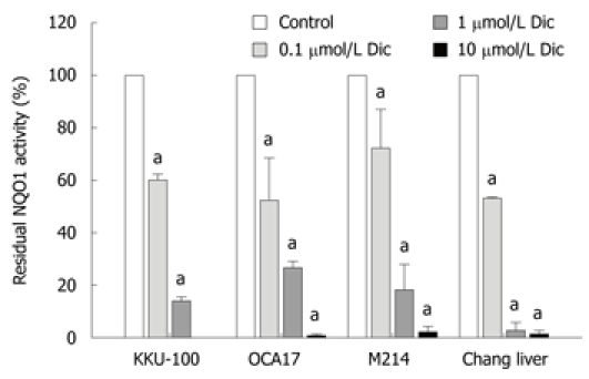
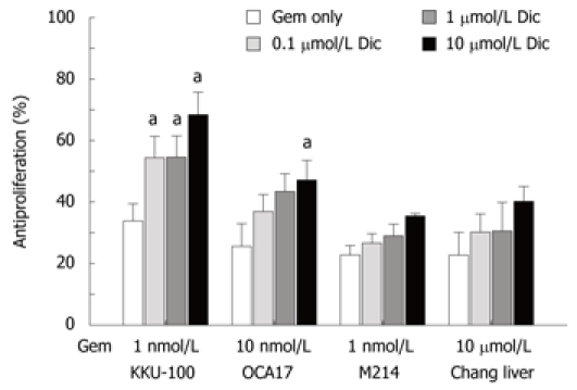
Aim: To investigate whether dicoumarol, a potent inhibitor of NAD(P)H quinone oxidoreductase-1 (NQO1), potentiates gemcitabine to induce cytotoxicity in cholangiocarcinoma cells (CCA) and the role of reactive oxygen generation in sensitizing the cells.

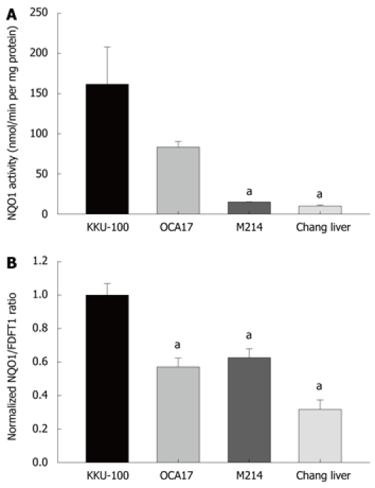
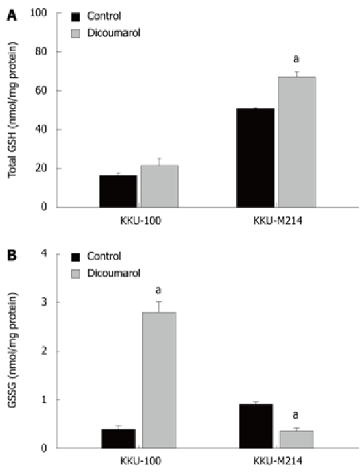
Methods: Four human cell lines with different NQO1 activity were used; the human CCA cell lines, KKU-100, KKU-OCA17, KKU-M214, and Chang liver cells. NQO1 activity and mRNA expression were determined. The cells were pretreated with dicoumarol at relevant concentrations before treatment with gemcitabine. Cytotoxicity was determined by staining with fluorescent dyes. Oxidant formation was examined by assay of cellular glutathione levels and reactive oxygen species production by using dihydrofluorescein diacetate. Measurement of mitochondrial transmembrane potential was performed by using JC-1 fluorescent probe. Western blotting analysis was performed to determine levels of survival related proteins.

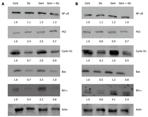
Results: Dicoumarol markedly enhanced the cytotoxicity of gemcitabine in KKU-100 and KKU-OCA17, the high NQO1 activity and mRNA expressing cells, but not in the other cells with low NQO1 activity. Dicoumarol induced a marked decrease in cellular redox of glutathione in KKU-100 cells, in contrast to KKU-M214 cells. Dicoumarol at concentrations that inhibited NQO1 activity did not alter mitochondrial transmembrane potential and production of reactive oxygen species. Gemcitabine alone induced activation of NF-kappaB and Bcl-(XL) protein expression. However, gemcitabine and dicoumarol combination induced increased p53 and decreased Bcl-(XL) levels in KKU-100, but not in KKU-M214 cells.

Conclusion: NQO1 may be important in sensitizing cells to anticancer drugs and inhibition of NQO1 may be a strategy for the treatment of CCA.


Figures

Figure 1
Figure 1
NQO1 activity and mRNA expression of cultured cells. A: NQO1 activity: Cholangiocarcinoma cells, KKU-100, KKU-OCA17, KKU-M214, and Chang liver cells were cultured in 96-well plates for assay of NQO1 activity by enzymatic methods; B: Expression of NQO1 mRNA: The cells were cultured in 6-well plates. Total RNA was extracted by Trizol reagent, converted to cDNA and analyzed by real-time PCR using FDFT1 as internal control. Bars represent mean ± SE, each from 3 experiments. aP < 0.05 vs control group.
Figure 2
Figure 2
Concentration response of NQO1 inhibition by dicoumarol. KKU-100, KKU-OCA17, KKU-M214, and Chang liver cells were cultured in 96-well plates overnight. The NQO1 activity was assayed in the presence of the inhibitor dicoumarol at concentrations from 0.1-10 μmol/L. Bars represent mean ± SE, each from 3 experiments. aP < 0.05 vs control group.
Figure 3
Figure 3
Potentiation of cytotoxicity of gemcitabine by dicoumarol. KKU-100, KKU-OCA17, KKU-M214, and Chang liver cells were cultured in 96-well plates overnight. Cultured cells were pretreated with dicoumarol (0.1-10 μmol/L) for 4 h before treatment with gemcitabine at 1 nmol/L, 10 nmol/L, 1 nmol/L and 10 μmol/L, for KKU-100, KKU-OCA17, KKU-M214, and Chang liver cells, respectively. Antiproliferation was analyzed by staining the cells with fluorescent dyes before examination under fluorescent microscope. Bars represent mean ± SE, each from 3 experiments. aP < 0.05 vs control group.
Figure 4
Figure 4
Glutathione redox status in cells treated with dicoumarol. KKU-100 and KKU-M214 cells were cultured and treated with dicoumarol (10 μmol/L) for 4 h, and cells were scraped for assays of (A) total glutathione and (B) glutathione disulfide. Bars represent mean ± SE, each from 3 experiments. aP < 0.05 vs control group.
Figure 5
Figure 5
Assay of mitochondrial transmembrane potential and reactive oxygen in CCA cells. The mitochondrial transmembrane potential was analyzed by using JC-1 fluorescent probe. Fluorescent readings of the J-aggregates and J monomers were used as measurement of mitochondrial transmembrane potential. KKU-100 and KKU-M214 cells were cultured in 96-well black plates. The cultured cells were pretreated with dicoumarol at 10 μmol/L for 4 h, then gemcitabine at 1 nmol/L was added and incubated at various times. A: Incubation for 6 h; B: Incubation for 24 h; C: Other cultured cells were treated with dicoumarol at 50 and 150 μmol/L for 3 h. Bars represent mean ± SE, each from triplicate assay. aP < 0.05 vs control group.
Figure 6
Figure 6
Western blotting analysis of proteins related to survival. KKU-100 and KKU-M214 cells were cultured overnight, pretreated with 10 μmol/L dicoumarol for 4 h before being treated with 1 nmol/L gemcitabine for 24 h. Cultured cells were collected for Western blotting analysis using β-actin as an internal control for equal protein loading. A: KKU-100 cells; B: KKU-M214 cells. Cont: Controls; Dic: Dicoumarol alone; Gem: Gemcitabine alone; Gem + Dic: Combination of gemcitabine and dicoumarol. Values were an average from two experiments of the target protein normalized with the internal control.

References

    1. Talalay P, Dinkova-Kostova AT. Role of nicotinamide quinone oxidoreductase 1 (NQO1) in protection against toxicity of electrophiles and reactive oxygen intermediates. Methods Enzymol. 2004;382:355–364. - PubMed
    1. Asher G, Tsvetkov P, Kahana C, Shaul Y. A mechanism of ubiquitin-independent proteasomal degradation of the tumor suppressors p53 and p73. Genes Dev. 2005;19:316–321. - PMC - PubMed
    1. Nioi P, Hayes JD. Contribution of NAD(P)H:quinone oxidoreductase 1 to protection against carcinogenesis, and regulation of its gene by the Nrf2 basic-region leucine zipper and the arylhydrocarbon receptor basic helix-loop-helix transcription factors. Mutat Res. 2004;555:149–171. - PubMed
    1. Siegel D, Gustafson DL, Dehn DL, Han JY, Boonchoong P, Berliner LJ, Ross D. NAD(P)H:quinone oxidoreductase 1: role as a superoxide scavenger. Mol Pharmacol. 2004;65:1238–1247. - PubMed
    1. Nair S, Xu C, Shen G, Hebbar V, Gopalakrishnan A, Hu R, Jain MR, Lin W, Keum YS, Liew C, et al. Pharmacogenomics of phenolic antioxidant butylated hydroxyanisole (BHA) in the small intestine and liver of Nrf2 knockout and C57BL/6J mice. Pharm Res. 2006;23:2621–2637. - PubMed

Publication types

MeSH terms