Review
. 2010 Jun 22;49(24):4945-56.
doi: 10.1021/bi1004798.

Human iron-sulfur cluster assembly, cellular iron homeostasis, and disease

Affiliations
Free PMC article
Review

Human iron-sulfur cluster assembly, cellular iron homeostasis, and disease

Hong Ye et al. Biochemistry. .
Free PMC article

Abstract

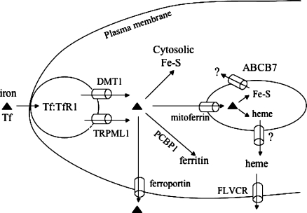
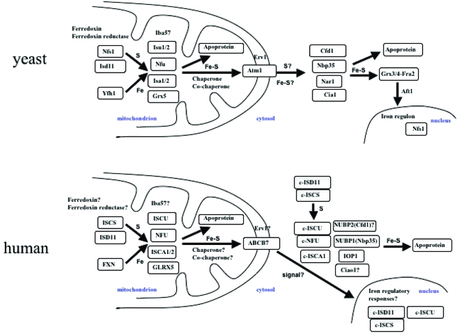
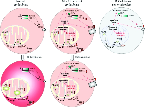
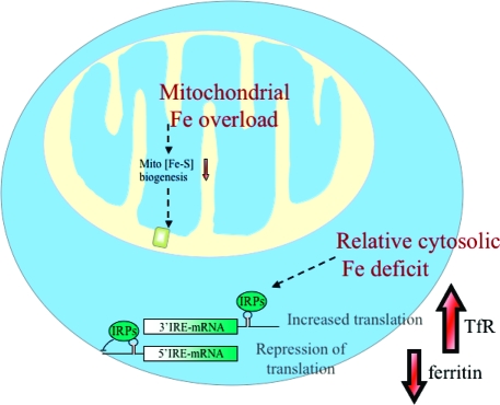
Iron-sulfur (Fe-S) proteins contain prosthetic groups consisting of two or more iron atoms bridged by sulfur ligands, which facilitate multiple functions, including redox activity, enzymatic function, and maintenance of structural integrity. More than 20 proteins are involved in the biosynthesis of iron-sulfur clusters in eukaryotes. Defective Fe-S cluster synthesis not only affects activities of many iron-sulfur enzymes, such as aconitase and succinate dehydrogenase, but also alters the regulation of cellular iron homeostasis, causing both mitochondrial iron overload and cytosolic iron deficiency. In this work, we review human Fe-S cluster biogenesis and human diseases that are caused by defective Fe-S cluster biogenesis. Fe-S cluster biogenesis takes place essentially in every tissue of humans, and products of human disease genes, including frataxin, GLRX5, ISCU, and ABCB7, have important roles in the process. However, the human diseases, Friedreich ataxia, glutaredoxin 5-deficient sideroblastic anemia, ISCU myopathy, and ABCB7 sideroblastic anemia/ataxia syndrome, affect specific tissues, while sparing others. Here we discuss the phenotypes caused by mutations in these different disease genes, and we compare the underlying pathophysiology and discuss the possible explanations for tissue-specific pathology in these diseases caused by defective Fe-S cluster biogenesis.


Figures

Figure 1
Figure 1
Subcellular iron trafficking in human cells. Iron is bound to transferrin Tf in serum, which interacts with TfR1 on the cell membrane. Formation of the Tf−TfR1 complex induces endocytosis. Upon acidification of the endosome, iron is released and then exported into the cytosol by DMT1 or TRPML1. Iron in cytosol has four main fates. It can be used to assemble Fe−S clusters and other iron proteins in cytosol, undergo transport into mitochondria via the mitochondrial iron importer, mitoferrin, be loaded into ferritin with the coordination of the PCBP1 chaperone, or be exported by ferroportin. Upon its import into mitochondria, iron is used to synthesize Fe−S clusters and heme. An unknown compound that represents the mitochondrial iron status and is a product of Fe−S cluster synthesis appears to be exported by ABCB7, which can affect cytosolic Fe−S cluster biogenesis. Heme is exported from the mitochondria by an unknown mechanism and is exported out of some cells by a heme transporter known as FLVCR.
Figure 2
Figure 2
Comparison of Fe−S cluster biogenesis pathways in eukaryotes. In yeast mitochondria, Nfs1 and Isd11 form a complex of cysteine desulfurase, which provides sulfur to scaffold proteins for Fe−S cluster assembly. The pair of ferredoxin (Yah1) and ferredoxin reductase (Arh1) perhaps provides reducing equivalents needed for cluster assembly. The yeast frataxin homologue, Yfh1, provides iron. Iron−sulfur clusters are assembled on scaffolds, and there are several alternative scaffolds, including Isu1 and -2, Nfu, Isa1 and -2, and Grx5. Iba57 is thought to function with the Isa1 and -2 scaffold proteins. Facilitated by chaperone (Ssq1) and cochaperone (Jac1) activities, the preassembled Fe−S clusters of Isu1 and -2 are delivered to target apoproteins in mitochondria. The mitochondrial inner membrane transporter Atm1 has been proposed to export either Fe−S cluster or sulfur to the cytosol, with assistance from Erv1 in the intermembrane space of mitochondria, and this compound “X” could then be used to assemble Fe−S clusters in the cytosol by scaffold proteins, including Cfd1, Nbp35, Nar1, and Cia1. The assembled Fe−S clusters are delivered to apoproteins, including the Grx3/4−Fra2 complex, which regulates the nucleocytoplasmic translocation of Aft1 by an unknown mechanism. As a transcription factor, Aft1 regulates transcription of the iron regulon in the yeast nucleus. Interestingly, there is a fraction of Nfs1 proteins present in yeast nucleus (109). In human mitochondria, ISCS and ISD11 form a complex that provides sulfur to scaffold proteins for Fe−S cluster biogenesis, while FXN is thought to provide iron. Potential scaffold proteins include ISCU, NFU, ISCA1/2, and GLRX5. The assembled Fe−S clusters are delivered to apoproteins for maturation. The roles of human ferredoxin, ferredoxin reductase, Iba57, chaperone, and cochaperone homologues in mitochondrial Fe−S cluster biogenesis remain to be confirmed. An unknown molecule that depends on mitochondrial Fe−S cluster biogenesis for function is exported by ABCB7. We propose that the ABCB7-exported molecule may serve as a signal that induces iron transcriptional remodeling in the nucleus to appropriately regulate mitochondrial iron homeostasis in response to a signal received from the mitochondria. In the cytosol, the cytosolic forms of ISCS and ISD11, c-ISCS and c-ISD11, respectively, provide sulfur, while iron can be directly acquired from cytosol, though its exact molecular source is not known. Fe−S clusters are likely synthesized de novo on c-ISCU, c-NFU, c-ISCA1, or NUBP1 (Nbp35) scaffolds. IOP1 may function in the delivery of the cluster to apoproteins. The involvement of the human Cfd1 homologue (NUBP2) and Cia1 homologue (Ciao1) in cytosolic Fe−S cluster biogenesis remains to be studied.
Figure 3
Figure 3
GLRX5 deficiency causes anemia but does not significantly affect non-erythroid tissues. In normal erythroblasts, ALAS2 and FECH in mitochondria contribute to heme synthesis, which is incorporated into hemoglobin (Hb). Erythroblasts are the only cells that express ALAS2, which contains a IRE in its 5′ UTR. All other cells express ALAS1, which is not regulated by the IRE−IRP system. Erythroblasts encode two forms of the ferroportin (FPN1) transcript, IRE-FPN1a and non-IRE-FPN1b. Both encode an identical FPN1 protein that functions as the iron exporter. Upon erythroid differentiation, both ALAS2 expression and FECH expression are upregulated, thus increasing the level of synthesis of heme and hemoglobin, while FPN1 expression is downregulated (65). In contrast, Fe−S cluster biogenesis is defective in GLRX5-deficient erythroblasts, iron accumulates in mitochondria, and associated iron deficiency in cytosol activates IRP proteins. The level of ALAS2 expression is decreased by IRP repression of its 5′ IRE. The level of FECH expression is also decreased, most likely because FECH does not acquire the Fe−S cluster it needs for stabilization (69). Together, heme synthesis and subsequent hemoglobinization are inhibited. Levels of expression of both FPN1 transcripts, including both FPN1a and FPN1b, are increased, perhaps in response to the stress caused by mitochondrial iron overload. Because FPN1b lacks the IRE, the expression of FPN1b evades the IRP repression and increases the level of expression of FPN1, which may exacerbate cytosolic iron deficiency. As a result, the GLRX5-deficient erythroblasts fail to produce enough heme for hemoglobinization upon differentiation. Although GLRX5-deficient non-erythroblasts also demonstrate iron overload in mitochondria and cytosolic iron deficiency, they express ALAS1, which does not have IRE and is not repressed by IRPs. Thus, heme synthesis is not significantly impaired in non-erythroid cell types. In addition, non-erythroblast cells express only FPN1a, which can be repressed by IRPs as cells develop cytosolic iron depletion.
Figure 4
Figure 4
Example of mitochondrial iron overload and its reversal when iron−sulfur cluster biogenesis is restored. Shown in the left panels are several fibroblasts that are stained with the Perls’ DAB iron staining technique, which detects ferric iron and enhances the signal with precipitation of DAB. Iron overload is present in a reticular pattern consistent with the mitochondrial network of patient fibroblast cells (left), but the iron deposits disappeared in patient cells rescued by the wild-type GLRX5 gene (right), introduced either by transfection (top) or by viral transduction (bottom) (32). The scale bars are 10 μm.
Figure 5
Figure 5
Deficiency in mitochondrial Fe−S cluster biogenesis causes mitochondrial iron overload, relative cytosolic iron deficiency, and activation of the cytosolic iron sensor proteins, IRP1 and IRP2. Deficiency in many of the major proteins involved in mitochondrial Fe−S cluster biogenesis results in defective mitochondrial Fe−S cluster biogenesis, which in turn results in transcriptional remodeling, mitochondrial iron overload, and cytosolic iron deficiency. IRP proteins are activated to be RNA regulatory proteins, which increase the rate of translation of 3′ IRE-mRNA, such as TfR1, whereas they inhibit the translation of mRNAs that contain IREs in the 5′ UTR, such as ferritin and ferroportin.

References

    1. Yin L.; Wu N.; Curtin J. C.; Qatanani M.; Szwergold N. R.; Reid R. A.; Waitt G. M.; Parks D. J.; Pearce K. H.; Wisely G. B.; Lazar M. A. (2007) Rev-erbα, a heme sensor that coordinates metabolic and circadian pathways. Science 318, 1786–1789. - PubMed
    1. Faller M.; Matsunaga M.; Yin S.; Loo J. A.; Guo F. (2007) Heme is involved in microRNA processing. Nat. Struct. Mol. Biol. 14, 23–29. - PubMed
    1. Hentze M. W.; Muckenthaler M. U.; Andrews N. C. (2004) Balancing acts: Molecular control of mammalian iron metabolism. Cell 117, 285–297. - PubMed
    1. De Domenico I.; McVey Ward D.; Kaplan J. (2008) Regulation of iron acquisition and storage: Consequences for iron-linked disorders. Nat. Rev. Mol. Cell Biol. 9, 72–81. - PubMed
    1. Ganz T. (2008) Iron homeostasis: Fitting the puzzle pieces together. Cell Metab. 7, 288–290. - PubMed

Publication types