. 2010 Jul 13;49(27):5817-27.
doi: 10.1021/bi100332r.

RNA internal loops with tandem AG pairs: the structure of the 5'GAGU/3'UGAG loop can be dramatically different from others, including 5'AAGU/3'UGAA

Affiliations
Free PMC article

RNA internal loops with tandem AG pairs: the structure of the 5'GAGU/3'UGAG loop can be dramatically different from others, including 5'AAGU/3'UGAA

Nicholas B Hammond et al. Biochemistry. .
Free PMC article

Abstract

Thermodynamic stabilities of 2 x 2 nucleotide tandem AG internal loops in RNA range from -1.3 to +3.4 kcal/mol at 37 degrees C and are not predicted well with a hydrogen-bonding model. To provide structural information to facilitate development of more sophisticated models for the sequence dependence of stability, we report the NMR solution structures of five RNA duplexes: (rGACGAGCGUCA)(2), (rGACUAGAGUCA)(2), (rGACAAGUGUCA)(2), (rGGUAGGCCA)(2), and (rGACGAGUGUCA)(2). The structures of these duplexes are compared to that of the previously solved (rGGCAGGCC)(2) (Wu, M., SantaLucia, J., Jr., and Turner, D. H. (1997) Biochemistry 36, 4449-4460). For loops bounded by Watson-Crick pairs, the AG and Watson-Crick pairs are all head-to-head imino-paired (cis Watson-Crick/Watson-Crick). The structures suggest that the sequence-dependent stability may reflect non-hydrogen-bonding interactions. Of the two loops bounded by G-U pairs, only the 5'UAGG/3'GGAU loop adopts canonical UG wobble pairing (cis Watson-Crick/Watson-Crick), with AG pairs that are only weakly imino-paired. Strikingly, the 5'GAGU/3'UGAG loop has two distinct duplex conformations, the major of which has both guanosine residues (G4 and G6 in (rGACGAGUGUCA)(2)) in a syn glycosidic bond conformation and forming a sheared GG pair (G4-G6*, GG trans Watson-Crick/Hoogsteen), both uracils (U7 and U7*) flipped out of the helix, and an AA pair (A5-A5*) in a dynamic or stacked conformation. These structures provide benchmarks for computational investigations into interactions responsible for the unexpected differences in loop free energies and structure.


Figures

Figure 1
Figure 1
1D NMR spectra of the imino proton regions at various temperatures for (rGACGAGCGUCA)2, (rGACAAGUGUCA)2, and (rGACUAGAGUCA)2 (left to right) at ∼2 mM strand concentration. Spectra were taken in 10 mM sodium phosphate buffer with 0.5 mM Na2EDTA and 80 mM NaCl at pH 6.1.
Figure 2
Figure 2
1D NMR spectra of the imino proton regions at various temperatures for 1 mM (rGACGAGUGUCA)2 (left, with 5′GABrGU/3′UBrGAG at bottom and 5′GAMeGU/3′UMeGAG second from bottom, both at 0 °C) and 1 mM (rGGUAGGCCA)2 (right). Spectra were taken in 10 mM sodium phosphate buffer with 0.5 mM Na2EDTA and 80 mM NaCl at pH 6.1.
Figure 3
Figure 3
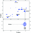
NOESY spectra of (rGACGAGCGUCA)2 in H2O at 1 °C with 150 ms mixing time (left and middle) and in D2O at 15 °C with 400 ms mixing time (right). The black line connects the G6 amino-H1 cross-peak to the G6 amino-A5H2 cross-peak in the same spectrum and then continues on to indicate the lack of a cross-peak in D2O, confirming the identity of the G6 amino-A5H2 cross-peak. Also of note is the A5H2-G6H1 cross-peak (left). These confirm the imino pairing in the AG loop (shown bottom, right) as these cross-peaks would not be seen in a sheared confirmation.
Figure 4
Figure 4
1D NMR spectra of the imino proton region of (rGACGAGUGUCA)2 at 0 °C at 0.66, 2, and 5 mM strand concentration (bottom to top). The size of the spectrum at 2 mM is reduced to allow easier comparisons to chemical shifts in the 5 mM spectrum.
Figure 5
Figure 5
2D NOESY spectra of (rGACGAGUGUCA)2 showing large H8-H1′ cross-peaks for G4 and G6 indicating syn conformation and G6H1-G4*H8 indicating G6-G4* pair.
Figure 6
Figure 6
Major groove view of the lowest energy structures with no NOE distance violations for (left to right) (rGACGAGCGUCA)2, (rGACAAGUGUCA)2, (rGACUAGAGUCA)2, and (rGGUAGGCCA)2 with the terminal base pairs and dangling adenosines removed. The loop adenosines and guanines are colored blue and red, respectively, while closing base pairs are in brown and all other base pairs are in black.
Figure 7
Figure 7
GG pair (trans Watson−Crick/Hoogsteen) found between G4 and G6* of (rGACGAGUGUCA)2 (5′GAGU/3′UGAG) (top) and schematic of the hydrogen-bonding network of the entire (rGACGAGUGUCA)2 duplex (bottom).
Figure 8
Figure 8
Top view of A4H2 (in red) in (rGACAAGUGUCA)2 (5′AAGU/3′UGAA) showing the proton stacked between the pyrimidine rings (yellow) of the 5′ cross-strand G6* (top) and the 3′ intrastrand A5 (bottom).
Figure 9
Figure 9
In the 5′GAGC/3′CGAG loop (left), viewed from the major groove, G4O6 (red) can be seen hydrogen bonding to the cross-strand amino of C7 (blue) and stacking above the amino of A5 (blue). Similarly, G6O6 has a pattern of hydrogen bonding to the A5 amino and stacking underneath an amino group. A similar pattern is observed for 5′UAGA/3′AGAU (second from right). When the sequence is changed to 5′CAGG/3′GGAC (second from left) or 5′AAGU/3′UGAA (right), this favorable electrostatic interaction is replaced with an unfavorable amino stacking on amino and oxygen stacking on oxygen interaction. The O-amino−O-amino interaction could add stability to 5′GAGC/3′CGAG and 5′UAGA/3′AGAU that is lost when the sequences are changed to 5′CAGG/3′GGAC and 5′AAGU/3′UGAA. The loop sequences and ΔG°loop are below their corresponding model.

Similar articles

Cited by

References

    1. Kruger K.; Grabowski P. J.; Zaug A. J.; Sands J.; Gottschling D. E.; Cech T. R. (1982) Self-splicing RNA: autoexcision and autocyclization of the ribosomal RNA intervening sequence of Tetrahymena. Cell 31, 147–157. - PubMed
    1. Guerrier-Takada C.; Gardiner K.; Marsh T.; Pace N.; Altman S. (1983) The RNA moiety of ribonuclease P is the catalytic subunit of the enzyme. Cell 35, 849–857. - PubMed
    1. Gesteland R. F., Cech T. R., and Atkins J. F. (2006) The RNA World, 3rd ed., Cold Spring Harbor Press, Cold Spring Harbor, NY.
    1. Kota J.; Chivukula R. R.; O’Donnell K. A.; Wentzel E. A.; Montgomery C. L.; Hwang H.; Chang T.; Vivekanandan P.; Torbenson M.; Clark K. R.; Mendell J. R.; Mendell J. T. (2009) Therapeutic microRNA delivery suppresses tumorigenesis in a murine liver cancer model. Cell 137, 1005–1017. - PMC - PubMed
    1. Sul J.; Wu C. K.; Jochems J.; Lee M. T.; Kim T. K.; Peritz T.; Buckley P.; Cappelleri D. J.; Maronski M.; Kim M.; Kumar V.; Meaney D.; Kim J.; Eberwine J. (2009) Transcriptome transfer produces a predictable cellular phenotype. Proc. Natl. Acad. Sci. U.S.A. 106, 7624–7629. - PMC - PubMed

Publication types