

A cephalometric study to investigate the skeletal relationships in patients with increasing severity of hypodontia
- PMID: 20482356
- PMCID: PMC8966445
- DOI: 10.2319/072309-411.1
A cephalometric study to investigate the skeletal relationships in patients with increasing severity of hypodontia
Abstract
Objectives: To determine the skeletal relationships in patients with hypodontia and analyze the effects of severity and pattern.
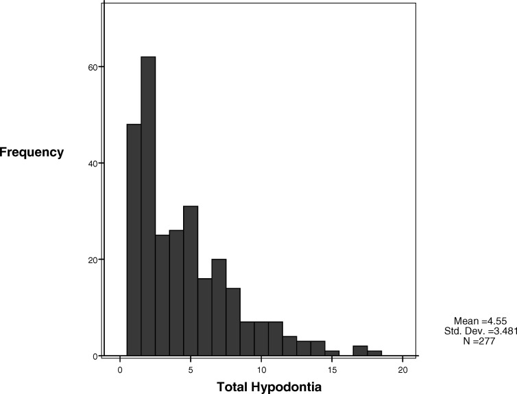
Materials and methods: Pretreatment lateral cephalograms from 277 patients with hypodontia, categorized by the number of missing teeth as mild (1-2), moderate (3-5), or severe (> or =6), were digitized recording angular measurements and ratios and compared with published norms matched for age and gender. Pattern was determined as mandibular, maxillary, bimaxillary, bilateral, anterior, posterior, and anteroposterior. Linear regression models assessed relationships between number of missing teeth and cephalometric parameters, controlling for the pattern of hypodontia.
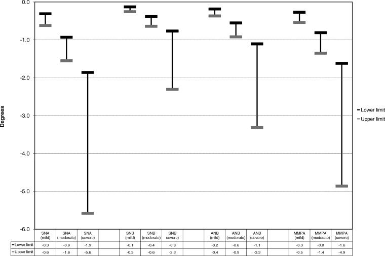
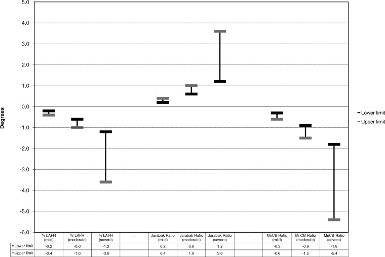
Results: For every additional missing tooth, SNA, SNB, and ANB decreased 0.3 degrees , 0.1 degrees , and 0.2 degrees , respectively; this was clinically significant for >4, >10, and >5 missing teeth, respectively. Mandibular to cranial base ratio decreased 0.3% for every additional missing tooth; this was clinically significant for >10 missing teeth. The MMPA decreased 0.3 degrees for every additional missing tooth; this was clinically significant for >7 missing teeth. Percentage LAFH decreased 0.2% for every additional missing tooth; this was significant for >7 missing teeth. Jarabak ratio increased 0.2% for each additional missing tooth; this was clinically significant for >10 missing teeth. Anterior hypodontia significantly decreased most cephalometric parameters.
Conclusions: Patients with hypodontia demonstrated a tendency toward a Class III relationship, caused by decreased maxillary and mandibular angular prognathism and MnCB ratio, though the effect was greater on the maxilla than the mandible. Clinical significance was only associated with severe hypodontia. Vertically, there was a tendency toward decreased MMPA and %LAFH; this was clinically relevant only with severe hypodontia. Anterior hypodontia had a significant effect on skeletal relationship.
Figures
Similar articles
-
Hypodontia patterns and variations in craniofacial morphology in Japanese orthodontic patients.Angle Orthod. 2006 Nov;76(6):996-1003. doi: 10.2319/082905-303. Angle Orthod. 2006. PMID: 17090161
-
Craniofacial profile in Southern Chinese with hypodontia.Eur J Orthod. 2009 Jun;31(3):300-5. doi: 10.1093/ejo/cjn111. Epub 2009 Feb 4. Eur J Orthod. 2009. PMID: 19193707
-
An analysis of the skeletal relationships in a group of young people with hypodontia.J Orthod. 2000 Dec;27(4):315-8. doi: 10.1093/ortho/27.4.315. J Orthod. 2000. PMID: 11099569
-
Craniofacial and dental features of Axenfeld-Rieger syndrome patients with PITX2 mutations.Orthod Craniofac Res. 2023 Aug;26(3):320-330. doi: 10.1111/ocr.12631. Epub 2023 Jan 18. Orthod Craniofac Res. 2023. PMID: 36620911 Review.
-
Congenitally missing teeth (hypodontia): A review of the literature concerning the etiology, prevalence, risk factors, patterns and treatment.Dent Res J (Isfahan). 2015 Jan-Feb;12(1):1-13. doi: 10.4103/1735-3327.150286. Dent Res J (Isfahan). 2015. PMID: 25709668 Free PMC article. Review.
Cited by
-
Dento-skeletal characteristics of cleft patients with missing teeth.Clin Cosmet Investig Dent. 2018 Nov 8;10:237-244. doi: 10.2147/CCIDE.S170717. eCollection 2018. Clin Cosmet Investig Dent. 2018. PMID: 30519115 Free PMC article.
-
Prevalence and patterns of permanent tooth agenesis in Down syndrome and their association with craniofacial morphology.Angle Orthod. 2011 Mar;81(2):260-9. doi: 10.2319/070910-391.1. Angle Orthod. 2011. PMID: 21208078 Free PMC article.
-
Association between Tooth Agenesis and Skeletal Malocclusions.J Oral Maxillofac Res. 2017 Jun 30;8(2):e3. doi: 10.5037/jomr.2017.8203. eCollection 2017 Apr-Jun. J Oral Maxillofac Res. 2017. PMID: 28791079 Free PMC article.
-
Prevalence and Pattern of Hypodontia among Croatian Orthodontic Patients.Acta Stomatol Croat. 2020 Jun;54(2):155-160. doi: 10.15644/asc54/2/5. Acta Stomatol Croat. 2020. PMID: 32801374 Free PMC article.
-
Comparison of craniofacial morphology in individuals with and without hypodontia with a special focus on the number of congenitally missing teeth.Front Public Health. 2022 Nov 18;10:1013862. doi: 10.3389/fpubh.2022.1013862. eCollection 2022. Front Public Health. 2022. PMID: 36466493 Free PMC article.
References
-
- Goodman J. R, Jones S. P, Hobkirk J. A, King P. A. Hypodontia: 1. Clinical features and the management of mild to moderate hypodontia. Dent Update. 1994;21:381–384.
-
- Dhanrajani P. J. Hypodontia: etiology, clinical features, and management. Quintessence Int. 2002;33:294–302. - PubMed
-
- Hobkirk J. A, Brook A. H. The management of patients with severe hypodontia. J Oral Rehabil. 1980;7:289–298. - PubMed
-
- Hobkirk J. A, King P. A, Goodman J. R, Jones S. P. Hypodontia: 2. The management of severe hypodontia. Dent Update. 1995;22:8–11. - PubMed
-
- Polder B. J, Van't Hof M. A, Van der Linden F. P. G. M, Kuijpers-Jagtman A. M. A meta-analysis of the prevalence of dental agenesis of permanent teeth. Community Dent Oral Epidemiol. 2004;32:217–226. - PubMed
MeSH terms
LinkOut - more resources
Full Text Sources
Research Materials
