. 2010;12(3):R93.
doi: 10.1186/ar3020. Epub 2010 May 18.

Treatment with apolipoprotein A-1 mimetic peptide reduces lupus-like manifestations in a murine lupus model of accelerated atherosclerosis

Affiliations

Treatment with apolipoprotein A-1 mimetic peptide reduces lupus-like manifestations in a murine lupus model of accelerated atherosclerosis

Jennifer M P Woo et al. Arthritis Res Ther. 2010.

Abstract

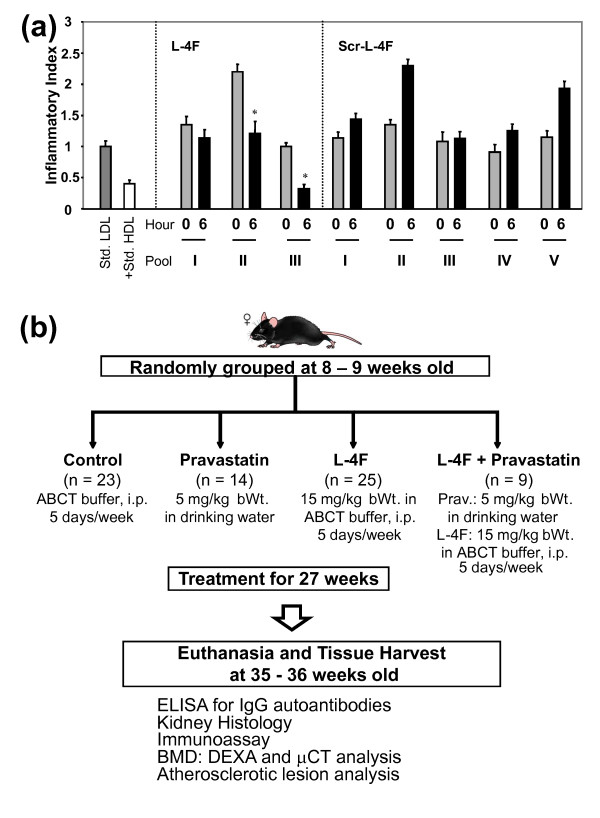
Introduction: The purpose of this study was to evaluate the effects of L-4F, an apolipoprotein A-1 mimetic peptide, alone or with pravastatin, in apoE-/-Fas-/-C57BL/6 mice that spontaneously develop immunoglobulin G (IgG) autoantibodies, glomerulonephritis, osteopenia, and atherosclerotic lesions on a normal chow diet.

Methods: Female mice, starting at eight to nine weeks of age, were treated for 27 weeks with 1) pravastatin, 2) L-4F, 3) L-4F plus pravastatin, or 4) vehicle control, followed by disease phenotype assessment.

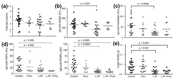
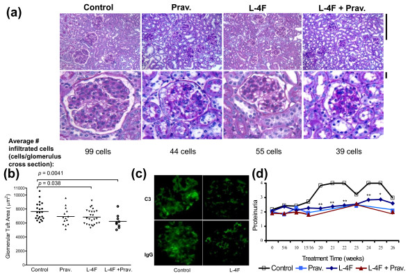
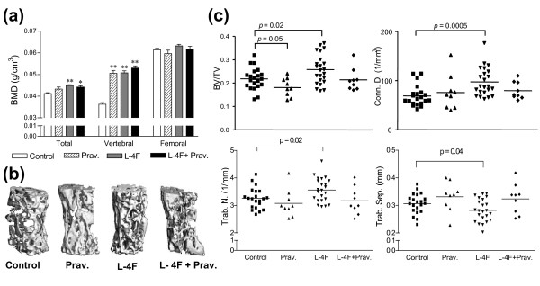
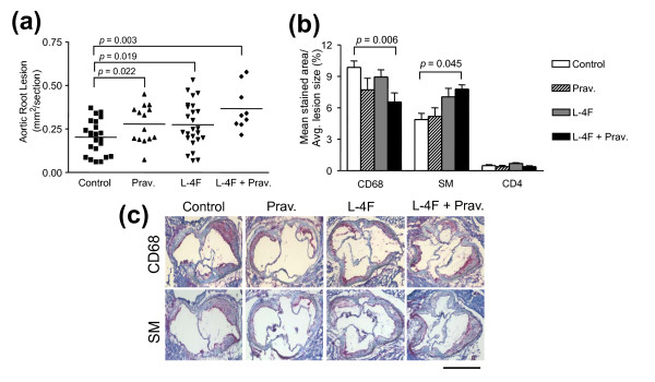
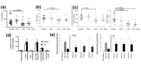
Results: In preliminary studies, dysfunctional, proinflammatory high-density lipoproteins (piHDL) were decreased six hours after a single L-4F, but not scrambled L-4F, injection in eight- to nine-week old mice. After 35 weeks, L-4F-treated mice, in the absence/presence of pravastatin, had significantly smaller lymph nodes and glomerular tufts (PL, LP<0.05), lower serum levels of IgG antibodies to double stranded DNA (dsDNA) (PL<0.05) and oxidized phospholipids (oxPLs) (PL, LP<0.005), and elevated total and vertebral bone mineral density (PL, LP<0.01) compared to vehicle controls. Although all treatment groups presented larger aortic root lesions compared to vehicle controls, enlarged atheromas in combination treatment mice had significantly less infiltrated CD68+ macrophages (PLP<0.01), significantly increased mean alpha-actin stained area (PLP<0.05), and significantly lower levels of circulating markers for atherosclerosis progression, CCL19 (PL, LP<0.0005) and VCAM-1 (PL<0.0002).

Conclusions: L-4F treatment, alone or with pravastatin, significantly reduced IgG anti-dsDNA and IgG anti-oxPLs, proteinuria, glomerulonephritis, and osteopenia in a murine lupus model of accelerated atherosclerosis. Despite enlarged aortic lesions, increased smooth muscle content, decreased macrophage infiltration, and decreased pro-atherogenic chemokines in L-4F plus pravastatin treated mice suggest protective mechanisms not only on lupus-like disease, but also on potential plaque remodeling in a murine model of systemic lupus erythematosus (SLE) and accelerated atherosclerosis.


Figures

Figure 1
Figure 1
Preliminary studies and experimental protocol. (a) Preliminary studies to determine the use of L-4F as a potential treatment in apoE-/-Fas-/- mice showed that HDL taken six hours after injection of L-4F was more successful in reducing LDL-induced monocyte chemotactic activity in cultures of human aortic endothelial cells compared to scrambled L-4F (Scr-L-4F). The value for No Addition (no LDL or HDL added to endothelial cultures) was subtracted from all values, the value for Std. LDL was taken as 1.0 and inflammatory index for LDL + HDL was calculated. Each pool represents HDL fractions from three to four mice. (b) Thirty-six week experimental protocol. *P ≤ 0.05.
Figure 2
Figure 2
Decreased auto-immune symptoms presented in mice treated with L-4F or combination treatment. ELISA assays on serum from randomly selected female apoE-/-Fas-/- mice at Week 35 or 36 showed: (a) comparable total serum IgG antibody levels among the different groups, suggesting an absence of general immune suppression, significantly reduced levels of (b) IgG anti-dsDNA and (c) IgG anti-cardiolipin in L-4F-treated mice, and (d) significantly lower IgG anti-PGPC and IgG anti-POVPC in mice treated with L-4F in the absence/presence of pravastatin. Pravastatin alone did not have any significant effect on IgG anti-dsDNA or IgG anti-oxPL levels. (e) In addition, lymph nodes from L-4F or L-4F plus pravastatin-treated mice were significantly smaller compared to control mice. Each symbol represents an individual mouse and the horizontal line represents the mean value. P-values < 0.05 were considered significant. AU, arbitrary units.
Figure 3
Figure 3
Improved renal lesions in female apoE-/-Fas-/- mice treated with L-4F or L-4F plus pravastatin. (a) Glomeruli of female mice treated with L-4F or L-4F plus pravastatin had smaller glomerular tufts compared to vehicle controls as seen in representative fields of renal cortex from each group (top panel; PAS stain; magnification ×400) and enlarged images from the corresponding field (bottom panel). Bars = 25 μm. In addition, the average number of infiltrated glomerular cells reflected this trend. (b) Quantification of glomerular tuft showed mice treated with L-4F or L-4F plus pravastatin had significantly decreased glomerular tuft area compared to vehicle controls (6,845 ± 1,060 and 6,226 ± 1,007 μm2 vs. 7,645 ± 1,200 μm2, respectively). (c) Immunofluorescence staining showed decreased deposition of IgG and C3 within kidneys of L-4F-treated mice compared to vehicle controls. (d) Starting Week 20 of treatment and through euthanasia, L-4F-treated mice had significantly lower levels of proteinuria compared to vehicle controls. *P ≤ 0.05; **P ≤ 0.01.
Figure 4
Figure 4
Increased bone mineral density and decreased osteopenia in L-4F and L-4F plus pravastatin treated mice. (a) Total and vertebral BMD (L2-L6), measured using DEXA, was increased in 35 to 36 week-old female apoE-/-Fas-/- mice when treated with pravastatin, L-4F, or in combination when compared to vehicle controls (total: 0.041 ± 0.002 vs. 0.043 ± 0.002 and 0.044 ± 0.002 and 0.044 ± 0.002 g/cm3, respectively and vertebral: 0.036 ± 0.004 vs. 0.051 ± 0.005 and 0.051 ± 0.005 and 0.053 ± 0.003 g/cm3, respectively). (b) μCT images of L5 lumbar vertebrae from female mice at 35 to 36 weeks of age. Mice treated with L-4F showed significant improvement in trabecular bone content. (c) Three-dimensional morphometric evaluation of L5 vertebrae. Mice treated with L-4F had significantly increased bone volume density (BV/TV), connectivity density (Conn. D.), and trabecular number (Trab. N.) and significantly lower trabecular separation (Trab. Sep.) when compared to controls. *P ≤ 0.01; **P ≤ 1E-07.
Figure 5
Figure 5
Evaluation of atherosclerotic manifestations. (a) Larger aortic lesions were seen in mice treated with pravastatin or L-4F or L-4F plus pravastatin when compared to vehicle controls (0.28 ± 0.11 and 0.27 ± 0.13 and 0.37 ± 0.13 μm2 vs. 0.19 ± 0.10 μm2, respectively). (b) Aortic lesions from L-4F plus pravastatin treated mice had significantly decreased macrophage infiltration when compared to vehicle controls (6.2 ± 1.2 vs. 9.8 ± 0.8%, respectively; P = 0.006). In addition, increased smooth muscle content in combination treatment mice compared to vehicle controls (7.8 ± 0.5% vs. 4.9 ± 2.3%, respectively; P = 0.04) suggests possible plaque remodeling. CD4+ T cell levels appeared unaltered by treatment. (c) Ten micrometer aortic root sections from female mice were stained for macrophage infiltration (CD68; rat anti-mouse CD68) and smooth muscle cells (SM, rat anti-mouse α-smooth muscle actin). Bar = 1 mm.
Figure 6
Figure 6
Unaffected lipid profiles with modified plasma antigen levels and monocyte chemotactic activity in representative mice. Luminex-based bead array was performed for plasma chemokines and cytokines, including: (a) IL-10 (interleukin-10; also known as human cytokine synthesis inhibitory factor, CSIF), a cytokine secreted in response to tissue damage, presented lower levels in L-4F-treated mice--consistent with increased tissue damage in control mice. (b) Plasma levels of CCL9 (also known as MIP-1γ), a chemoattractant that contributes to monocyte infiltration in renal disease, were significantly less in mice treated with L-4F. (c) Indicators of atherosclerosis severity: CCL19 (also known as MIP-3-β) and VCAM-1. CCL19 recruits T-cells and dendritic cells to target organs and promotes inflammatory responses and was significantly decreased in mice treated with L-4F or combination treatment. Similar trends were seen with VCAM-1, an endothelial adhesion molecule involved in atherosclerotic plaque formation and progression of glomerulonephritis. After Bonferoni correction, P-values less than 0.0009 for plasma markers were considered significant. (d) Plasma lipid levels, including total cholesterol, HDL cholesterol, and non-HDL cholesterol, were unaffected in all of the treatment groups compared to vehicle controls. (e) However, L-4F (L) significantly rendered mouse HDL anti-inflammatory and LDL less inflammatory compared to control (C) as determined in cultures of human aortic endothelial cells (n = 10 mice per treatment group, three to four mice per pool). *P ≤ 0.05.

Comment in

References

    1. Esdaile JM, Abrahamowicz M, Grodzicky T, Li Y, Panaritis C, du Berger R, Côte R, Grover SA, Fortin PR, Clarke AE, Senécal JL. Traditional Framingham risk factors fail to fully account for accelerated atherosclerosis in systemic lupus erythematosus. Arthritis and Rheumatism. 2001;44:2331–2337. doi: 10.1002/1529-0131(200110)44:10<2331::AID-ART395>3.0.CO;2-I. - DOI - PubMed
    1. Manzi S, Meilahn EN, Rairie JE, Conte CG, Medsger TA Jr, Jansen-McWilliams L, D'Agostino RB, Kuller LH. Age-specific incidence rates of myocardial infarction and angina in women with systemic lupus erythematosus: comparison with the Framingham Study. Am J Epidemiol. 1997;145:408–415. - PubMed
    1. Turesson C, Jacobsson LT, Matteson EL. Cardiovascular co-morbidity in rheumatic diseases. Vasc Health Risk Manag. 2008;4:605–614. - PMC - PubMed
    1. Roman MJ, Shanker BA, Davis A, Lockshin MD, Sammaritano L, Simantov R, Crow MK, Schwartz JE, Paget SA, Devereux RB, Salmon JE. Prevalence and correlates of accelerated atherosclerosis in systemic lupus erythematosus. N Engl J Med. 2003;349:2399–2406. doi: 10.1056/NEJMoa035471. - DOI - PubMed
    1. Asanuma Y, Oeser A, Shintani AK, Turner E, Olsen N, Fazio S, Linton MF, Raggi P, Stein CM. Premature coronary-artery atherosclerosis in systemic lupus erythematosus. N Engl J Med. 2003;349:2407–2415. doi: 10.1056/NEJMoa035611. - DOI - PubMed

Publication types

MeSH terms