. 2010 May 18:10:52.
doi: 10.1186/1471-213X-10-52.

Emergence and migration of trunk neural crest cells in a snake, the California Kingsnake (Lampropeltis getula californiae)

Affiliations

Emergence and migration of trunk neural crest cells in a snake, the California Kingsnake (Lampropeltis getula californiae)

Michelle Reyes et al. BMC Dev Biol. .

Abstract

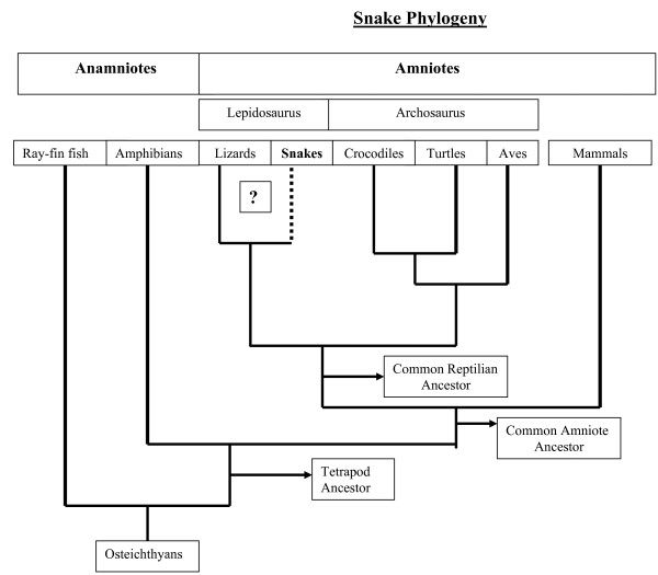
Background: The neural crest is a group of multipotent cells that emerges after an epithelial-to-mesenchymal transition from the dorsal neural tube early during development. These cells then migrate throughout the embryo, giving rise to a wide variety derivatives including the peripheral nervous system, craniofacial skeleton, pigment cells, and endocrine organs. While much is known about neural crest cells in mammals, birds, amphibians and fish, relatively little is known about their development in non-avian reptiles like snakes and lizards.

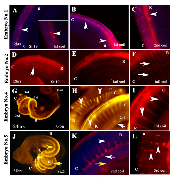
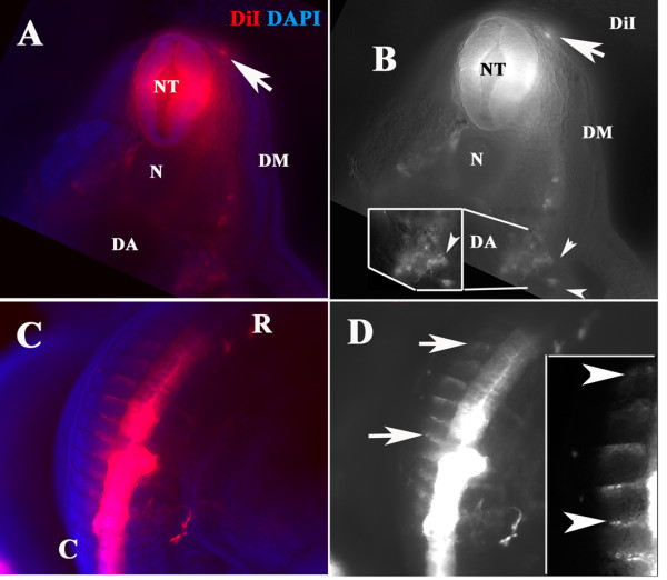
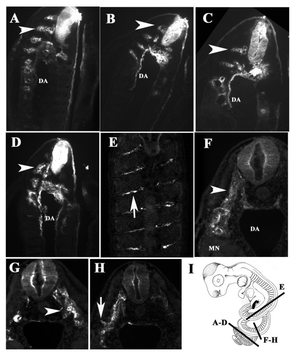
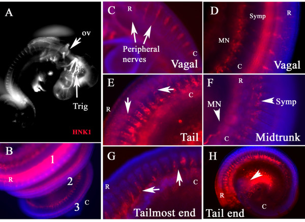
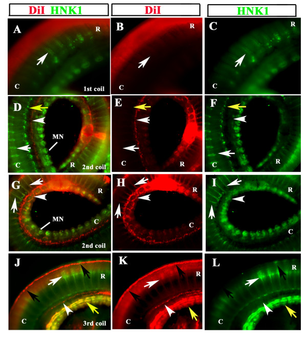
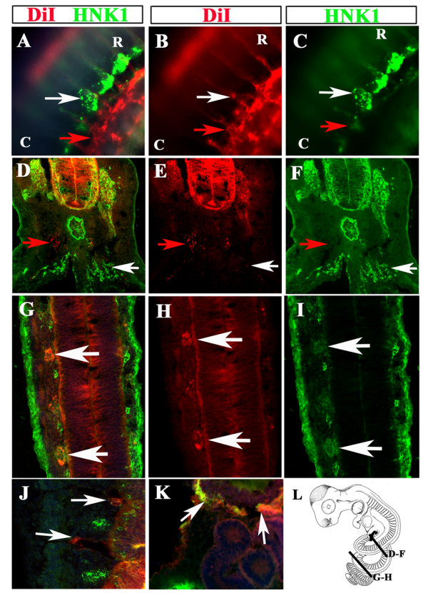
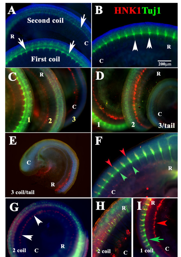
Results: In this study, we show for the first time ever trunk neural crest migration in a snake by labeling it with DiI and immunofluorescence. As in birds and mammals, we find that early migrating trunk neural crest cells use both a ventromedial pathway and an inter-somitic pathway in the snake. However, unlike birds and mammals, we also observed large numbers of late migrating neural crest cells utilizing the inter-somitic pathway in snake.

Conclusions: We found that while trunk neural crest migration in snakes is very similar to that of other amniotes, the inter-somitic pathway is used more extensively by late-migrating trunk neural crest cells in snake.


Figures

Figure 1
Figure 1
Phylogeny of Snake. We describe the phylogenetic position of snakes and other reptilians according to the most recent consensus [18]. The box with question marks refers to organisms for which neural crest migration has not been reported. The figure was drawn by the author, based upon Shedlock's (2007) recent phylogenetic analysis
Figure 2
Figure 2
Snake DiI injection. Snake embryos (1,2,4 and 5) were removed from the egg and the neural tube injected with DiI. After 12 hrs (A-F) or 24 hrs (G-L) of incubation embryos were fixed and photographed for migrating DiI-positive neural crest cells. Each embryo is shown in sequential higher magnifications. Embryo 1 show delaminated neural crest cells (arrowheads A-C), although it did not have substantial number of crest cells migrating in streams (higher magnification in panels B, C). Embryo 2 had clear robust migrating neural crest cells in streams (arrowhead in D for first coil). At the tail level the cells migrated on the rostral portion of the somites, further magnified in panel F. Embryos 4 and 5 showed a wider range of DiI labeling since it was incubated for 24 hrs and DiI labeling reached the tail end (G-L). Embryo 4 neural crest cells migrated as well as streams on the rostral side of somites (arrowheads in H and I) both in 2nd and 3rd coils as well as into the mesonephroi (arrows in H). Embryo 5 showed the rostrally migrating cells, in the 2nd coil we observe that cells were migrating between the somites (arrowhead in K) as well as cells along what is the presumptive aorta (arrow in K), also as for Embryo 4, DiI cells reached the mesonephroi (yellow arrows in J, K). L showed higher magnification of 3rd more caudal coil with individual cells migrating on the rostral portion of the somites (arrowheads in L). R is for rostral; C is for caudal orientation in the embryo coils.
Figure 3
Figure 3
Migrating cells travel same pathways as neural crest cells. A and B: Thick section of a DiI (red) and DAPI (blue) labeled snake embryo (Embryo 2 incubated for 12 hrs) on the second coil showed cells that migrated along the ventromedial pathway and settled by the dorsal aorta (D, A). Insert in B shows cells in more detail by the aorta. There was also a single DiI cell migrating sub-ectodermally (arrow in A, B). C and D show DiI (red) and DAPI (blue) labeled cells migrating on the most rostral portion of the somite (arrows in D point to area of the insert in D at higher magnification). This embryo (3 days POP) was labeled with DiI and incubated for 12 hrs at 37°C. N: notochord, DM: dermomyotome, NT: neural tube. R is for rostral; C is for caudal orientation in the embryo coils.
Figure 4
Figure 4
Sections through DiI labeled snake embryo. Snake embryos (Embryo 3 and 4) were cryo-sectioned. A-E shows sections starting at the most caudal portion and ending at the most rostral portion of st.20 Embryo 3. Neural crest cells showed robust migration in the tail region (arrowheads in A-C) and at more rostral levels the numbers were fewer and cells were found along the inter-somitic space (arrow in E). F-H show a transverse section close to the tail, showing thick streams of neural crest migrating ventromedially (F, G) or distally as if beginning to enter the sub-ectodermal space (arrow in H). I Cartoon depicting the axial location and orientation of these sections. DA: dorsal aorta. MN: mesonephroi. R is for rostral; C is for caudal orientation in the embryo coils.
Figure 5
Figure 5
Wholemount immuno-staining with HNK1. Snake embryo at st.21 (4POV) was wholemounted with HNK1 antibody (red) and DAPI (blue). A and B show head and trunk coils respectively. C and D show sensory ganglia positive for HNK1 at the vagal level. Arrows point spinal nerves positive for HNK1. E and F show HNK1 cells migrating along ventromedial pathway (arrows in E) at tail portion and crest cells at the sympathetic/aorta level or mesonephroi (arrowheads in F) at the midtrunk portion. G corresponds to the tail-most end, where the first neural crest cells are migrating (arrows). H shows the tail end providing a lower magnification of E and G areas. MN: mesonephroi. Symp: sympathetic. R corresponds to rostral and C to caudal.
Figure 6
Figure 6
DiI and HNK1 labels neural crest cells. DiI (red) labeled st.21 snake embryo No.5 was double stained with HNK1 (green). A-C shows 1st coil; D-I 2nd coil, with D_F more rostral versus the G-I more caudal portion; and J-L the 3rd coil. Each column shows either double channel, or just DiI or HNK1 staining. A-C DiI labeling of migrating crest was minimal, HNK1 faintly labeled some cells on the ventral portion of the trunk (white arrow). D-F shows strong DiI staining just above (white arrowhead) and in the mesonephroi, the point of injection in this embryo is marked as a large DiI spot. HNK1 staining labeled condensing peripheral ganglia, (white arrow), sympathetic ganglia (yellow arrow) and mesonephroi. G-H shows cells along the ventromedial pathway that are positive for both DiI and HNK1 (white arrows). DiI labeling also marked cells that now reached mesonephroi and are populating this tissue around (white arrowhead). J-L shows DiI-labeled cells that just delaminated from the neural tube but have not migrated (black arrows) as well as a small group of cells above the mesonephroi (white arrowhead). HNK1 labeled cells moving along the ventromedial pathway (white arrow) as well as the developing mesonephroi (yellow arrow). (The tail-most end of this embryo did not have DiI positive neural tube or migrating cells). R is for rostral; C is for caudal orientation in the embryo coils.
Figure 7
Figure 7
DiI and HNK1 labels neural crest cells. DiI (red) and HNK1 (green) st.21 stained snake embryo No.5 sections labels a variety of migrating neural crest. A-C show wholemount at the level of second coil, which label DiI migrating cells co-localizing with HNK1 around aorta/ventral portion of trunk (white arrows). There were many DiI cells around the mesonephroi (red arrows). D-F show a section on the caudal portion of 1st coil. The only DiI cells were localized in the DRG, which also was strongly stained with HNK1. Other cells were only DiI (red arrows) or only HNK1 (white arrows). G-I shows a longitudinal section on the second coil where DiI and HNK1 co-localized, especially in the condensing DRG. Not all DiI were HNK1 positive and vice-versa. J shows a higher magnification of one longitudinal section of DiI positive neural crest migrating between the somites (arrows) while HNK1 cells are found in the rostral portion of the somite. K corresponds to a section on the 2nd coil showing DiI co-localizing with HNK1 around the mesonephroi/kidneys (re.Fig. 5G-I). L is a cartoon depicting the axial levels of the sections shown in this figure. R is for rostral; C is for caudal orientation in the embryo coils.
Figure 8
Figure 8
Peripheral ganglia development in snake embryos. Wholemount staining of snake embryo with Tuj1 (green in A and B) or double stained with Tuj1 (green) and HNK1 (red) (C-I). DRG development progresses in a rostral to caudal manner: cervical DRGs are clearly mature (arrowheads in B) while those in the second coil and tail are not positive for Tuj1 (arrows in A and E). Only at more rostral levels we observe Tuj1 and HNK1 overlap (red and green arrowheads in F). G shows second coil where Tuj1 exhibited a spotted pattern, while HNK1 labeled the cells around the aorta as in other figures (arrowheads). H shows a higher magnification of G. I show a higher magnification of first coil. Blue stain corresponds to DAPI labeled nuclei. R is for rostral; C is for caudal orientation in the embryo coils.
Figure 9
Figure 9
Peripheral ganglia comparison among archosaurs. Snake st.21 (A), lizard (B), turtle (C) and chicken HH20 (D) and were wholemount stained for Tuj1. Cervical sensory ganglia in snake, lizard and turtle are spindle shaped, while chicken ganglia are triangular (arrowheads). Dotted lines outline the shape of the sensory ganglia. R is for rostral; C is for caudal orientation in the embryo coils.

Similar articles

Cited by

References

    1. Hall BK. The Neural Crest and Neural Crest Cells in Vertebrate Development and Evolution. New York: Springer-Verlag; 2009.
    1. Baker CV. Neural Crest and Cranial Ectodermal Placodes. 4. New York: Springer, New York; 2005.
    1. Freitas R, Zhang G, Albert JS, Evans DH, Cohn MJ. Developmental origin of shark electrosensory organs. Evol Dev. 2006;8:74–80. doi: 10.1111/j.1525-142X.2006.05076.x. - DOI - PubMed
    1. Smith M, Hickman A, Amanzee D, Lumsden A, Thorogood P. Trunk neural crest origin of caudal fin mesenchyme in the zebrafish Danio rerio. Proc R Soc Lond B Biol Sci. 1994;256:137–145. doi: 10.1098/rspb.1994.0061. - DOI
    1. Serbedzija GN, Bronner-Fraser M, Fraser SE. A vital dye analysis of the timing and pathways of avian trunk neural crest cell migration. Development. 1989;106:809–16. - PubMed

Publication types