. 2010 May 18:7:23.
doi: 10.1186/1476-9255-7-23.

Differential splicing of the apoptosis-associated speck like protein containing a caspase recruitment domain (ASC) regulates inflammasomes

Affiliations

Differential splicing of the apoptosis-associated speck like protein containing a caspase recruitment domain (ASC) regulates inflammasomes

Nicole B Bryan et al. J Inflamm (Lond). .

Abstract

Background: The apoptotic speck-like protein containing a caspase recruitment domain (ASC) is the essential adaptor protein for caspase 1 mediated interleukin (IL)-1beta and IL-18 processing in inflammasomes. It bridges activated Nod like receptors (NLRs), which are a family of cytosolic pattern recognition receptors of the innate immune system, with caspase 1, resulting in caspase 1 activation and subsequent processing of caspase 1 substrates. Hence, macrophages from ASC deficient mice are impaired in their ability to produce bioactive IL-1beta. Furthermore, we recently showed that ASC translocates from the nucleus to the cytosol in response to inflammatory stimulation in order to promote an inflammasome response, which triggers IL-1beta processing and secretion. However, the precise regulation of inflammasomes at the level of ASC is still not completely understood. In this study we identified and characterized three novel ASC isoforms for their ability to function as an inflammasome adaptor.

Methods: To establish the ability of ASC and ASC isoforms as functional inflammasome adaptors, IL-1beta processing and secretion was investigated by ELISA in inflammasome reconstitution assays, stable expression in THP-1 and J774A1 cells, and by restoring the lack of endogenous ASC in mouse RAW264.7 macrophages. In addition, the localization of ASC and ASC isoforms was determined by immunofluorescence staining.

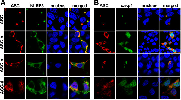
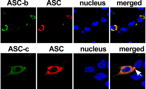
Results: The three novel ASC isoforms, ASC-b, ASC-c and ASC-d display unique and distinct capabilities to each other and to full length ASC in respect to their function as an inflammasome adaptor, with one of the isoforms even showing an inhibitory effect. Consistently, only the activating isoforms of ASC, ASC and ASC-b, co-localized with NLRP3 and caspase 1, while the inhibitory isoform ASC-c, co-localized only with caspase 1, but not with NLRP3. ASC-d did not co-localize with NLRP3 or with caspase 1 and consistently lacked the ability to function as an inflammasome adaptor and its precise function and relation to ASC will need further investigation.

Conclusions: Alternative splicing and potentially other editing mechanisms generate ASC isoforms with distinct abilities to function as inflammasome adaptor, which is potentially utilized to regulate inflammasomes during the inflammatory host response.


Figures

Figure 1
Figure 1
Identification of ASC isoforms. (A) Differentiated THP-1 macrophages were separated into nuclear and cytosolic fractions and analyzed for ASC expression using a monoclonal anti-ASC antibody recognizing the PYD of ASC by immunoblot. Blots were stripped and re-probed with antibodies for the cytosolic GAPDH and nuclear Lamin A to control for fractionation efficiency. (B) THP-1 lysates were analyzed by immunoblot for ASC expression using antibodies recognizing the PYD, the linker, and the CARD, respectively. (C) Lysates from PMA-differentiated and LPS-treated (300 ng/ml) THP-1 cells and J774A1 cells were separated by SDS/PAGE and immunoblotted with a PYD-specific anti-ASC antibody (AL177). (D) Lysates of human peripheral blood macrophages (PBM) that were left untreated, or treated with LPS for the indicated times, were immunoblotted for ASC. (E) PMA-differentiated THP-1 cells were treated with LPS (300 ng/ml) for the indicated times and analyzed by RT-PCR for ASC transcripts using the primer pairs pr-1 (ASC, 299 bp; ASC-b, 242 bp), pr-2 (ASC-c, 66 bp), and pr-3 (ASC and ASC-b, 128 bp; ASC-d, 100 bp). A short exposure (upper panel) and long exposure (middle panel) is shown, because of the relative low abundance of ASC-d transcripts. A β -actin primer pair (533 bp, lower panel) was used as a control.
Figure 2
Figure 2
Three novel ASC isoforms. (A) Clustal W alignment of ASC, ASC-b, ASC-c and ASC-d. ASC consists of a PYD, linker, and CARD, while ASC-b displays an in frame deletion of amino acids 93 to 111, corresponding to the complete linker region. ASC-c lacks amino acids 26 to 85 corresponding to helices 3 to 6 of the ASC-PYD, and in ASC-d amino acids 36-195 are replaced with 69 unrelated amino acids due to a frame shift resulting in the deletion of nucleotides 107 to 134 in ASC-d. (B) Schemata showing the domain structure of the ASC isoforms. (C) Myc-tagged ASC, ASC-b, ASC-c and ASC-d were transiently transfected into HEK293 cells and expression of ASC proteins with the predicted molecular weight was verified by immunoblot using anti-myc antibodies.
Figure 3
Figure 3
Localization of ASC isoforms. Subcellular localization of the myc-tagged ASC isoforms was examined in transiently transfected HEK293 cells. Cells were fixed and immunostained with monoclonal anti-myc antibodies and Alexa Fluor 488-conjugatetd secondary antibodies. Nuclei and actin were visualized using Topro-3 and Alexa Fluor 546-conjugated phalloidin, respectively. Images were acquired by laser scanning confocal microscopy, showing from left to right ASC (green), nucleus (blue), actin (red) and a merged composite image. The panels show ASC (1st and 2nd panels), ASC-b (3rd panel), ASC-c (4th panel), ASC-d (5th panel), and vector control (6th panel).
Figure 4
Figure 4
Localization of ASC isoforms, NLRP3, and caspase 1. ASC isoforms were transiently co-transfected into HEK293 cells with GFP-tagged NALP3R260W (A) or GFP-tagged pro-caspase 1C285A (B). Cells were fixed and immunostained with polyclonal anti-myc (Santa Cruz Biotechnology) and Alexa Fluor 546-conjugated secondary antibodies (Invitrogen). Topro-3 was used to visualize the nucleus. All images were acquired using laser scanning confocal microscopy with a 100x oil-immersion objective. Panels from left to right show ASC (red), NLRP3 or pro-caspase-1 (green), nucleus (blue), and a merged composite image.
Figure 5
Figure 5
Co-localization of ASC with ASC-b and ASC-c. HEK 293 cells were transiently co-transfected with HA-tagged ASC and myc-tagged ASC-b (1st panel) or ASC-c (2nd panel). Cells were fixed and immunostained with monoclonal anti-myc (Millipore) and polyclonal anti-HA (Abcam) antibodies, and Alexa Fluor-488 and -546 conjugated secondary antibodies (Invitrogen), respectively. Topro-3 was used to visualize the nucleus. All images were acquired using laser scanning confocal microscopy with a 100× oil-immersion objective. Panels from left to right show ASC-b/ASC-c (green), ASC (red), nucleus (blue) and a merged composite image. An arrow points to the aggregate.
Figure 6
Figure 6
Distinct ASC isoforms can function as either activating or inhibitory inflammasome adaptor. (A) Inflammasomes were reconstituted in HEK293 cells by transient transfection of pro-IL-1β, pro-caspase 1, ASC, ASC-b, ASC-c, ASC-d, in the absence (black bars) or presence (gray bars) of the constitutive active NLRP3R260W, as indicated. Culture supernatants were analyzed for secreted IL-1β by ELISA 36 hours post transfection. (B) The ASC deficient RAW264.7 mouse macrophage cell line was stably transfected with empty vector, myc-tagged ASC, or myc-tagged ASC-b and analyzed for IL-1β release in resting cells (black bars) and following LPS (300 ng/ml)/ATP (5 mM) activation (gray bars). (C) Inflammasomes were reconstituted in HEK293 cells as shown in Figure 6A. Secreted IL-1β was analyzed by ELISA. All experiments were performed in triplicates (n = 3, +/- SD). (D) Control THP-1 cells (Ctrl) or THP-1 cells stably expressing high levels of ASC-c (#1) or low levels of ASC-c (#2) were treated with LPS (300 ng/ml) for 16 hours and analyzed for IL-1β release. Expression of ASC-c was determined by immunoblot. (E) Control J774A1 cells (Ctrl) or J774A1 cells stably expressing ASC-c were treated with LPS (300 ng/ml) for 16 hours, pulsed with 3 mM ATP for 15 minutes and analyzed for IL-1β release. Experiments in D and E were performed in triplicates (n = 2, +/- SD). Expression of ASC-c was determined by immunoblot. Note that the lysates from THP-1 and J774A1 cells were separated on the same gel and are the same exposure time.
Figure 7
Figure 7
ASC and ASC-b interact with pro-caspase 1 with similar affinity. Immobilized GST-caspase 1-CARD was incubated with in vitro translated and biotinylated ASC or ASC-b and subjected to in vitro GST-pull down assays using GST-caspase 1-CARD and GST control immobilized to GSH Sepharose, as indicated. Bound proteins were visualized with streptavidin-HRP and ECL-Plus detection (Amersham Pharmacia Biotech). 10% of the in vitro translated proteins were loaded as 'input".

References

    1. Martinon F, Burns K, Tschopp J. The Inflammasome: A molecular platform triggering activation of inflammatory caspases and processing of proIL-1b. Mol Cell. 2002;10:417–426. doi: 10.1016/S1097-2765(02)00599-3. - DOI - PubMed
    1. Dinarello CA. Interleukin-1 beta, interleukin-18, and the interleukin-1 beta converting enzyme. Ann N Y Acad Sci. 1998;856:1–11. doi: 10.1111/j.1749-6632.1998.tb08307.x. - DOI - PubMed
    1. Keller M, Ruegg A, Werner S, Beer HD. Active caspase 1 is a regulator of unconventional protein secretion. Cell. 2008;132:818–831. doi: 10.1016/j.cell.2007.12.040. - DOI - PubMed
    1. Shao W, Yeretssian G, Doiron K, Hussain SN, Saleh M. The caspase 1 digestome identifies the glycolysis pathway as a target during infection and septic shock. J Biol Chem. 2007;282:36321–36329. doi: 10.1074/jbc.M708182200. - DOI - PubMed
    1. Dinarello CA. Immunological and inflammatory functions of the interleukin-1 family. Annu Rev Immunol. 2009;27:519–550. doi: 10.1146/annurev.immunol.021908.132612. - DOI - PubMed