. 2010 May 19:9:112.
doi: 10.1186/1476-4598-9-112.

Pristimerin induces apoptosis in imatinib-resistant chronic myelogenous leukemia cells harboring T315I mutation by blocking NF-kappaB signaling and depleting Bcr-Abl

Affiliations

Pristimerin induces apoptosis in imatinib-resistant chronic myelogenous leukemia cells harboring T315I mutation by blocking NF-kappaB signaling and depleting Bcr-Abl

Zhongzheng Lu et al. Mol Cancer. .

Erratum in

Abstract

Background: Chronic myelogenous leukemia (CML) is characterized by the chimeric tyrosine kinase Bcr-Abl. Bcr-Abl-T315I is the notorious point mutation that causes resistance to imatinib and the second generation tyrosine kinase inhibitors, leading to poor prognosis. CML blasts have constitutive p65 (RelA NF-kappaB) transcriptional activity, and NF-kappaB may be a potential target for molecular therapies in CML that may also be effective against CML cells with Bcr-Abl-T315I.

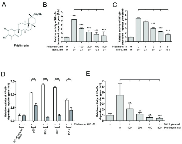
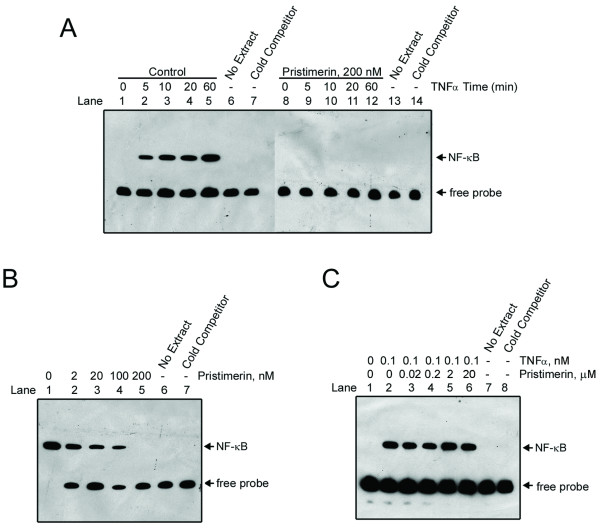
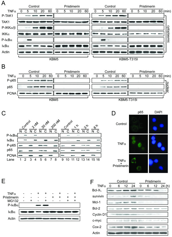
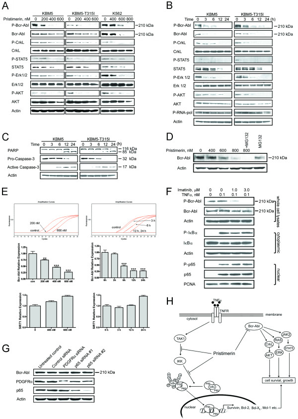
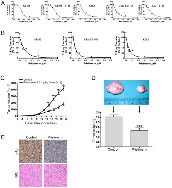
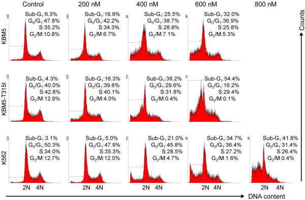
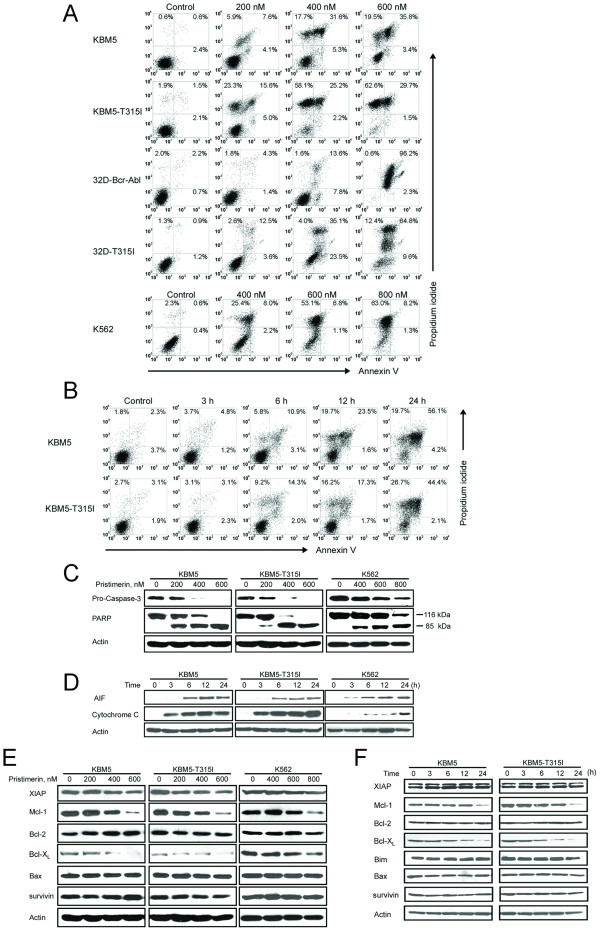
Results: In this report, we discovered that pristimerin, a quinonemethide triterpenoid isolated from Celastraceae and Hippocrateaceae, inhibited growth and induced apoptosis in CML cells, including the cells harboring Bcr-Abl-T315I mutation. Additionally, pristimerin inhibited the growth of imatinib-resistant Bcr-Abl-T315I xenografts in nude mice. Pristimerin blocked the TNFalpha-induced IkappaBalpha phosphorylation, translocation of p65, and expression of NF-kappaB-regulated genes. Pristimerin inhibited two steps in NF-kappaB signaling: TAK1TauIKK and IKKTauIkappaBalpha. Pristimerin potently inhibited two pairs of CML cell lines (KBM5 versus KBM5-T315I, 32D-Bcr-Abl versus 32D-Bcr-Abl-T315I) and primary cells from a CML patient with acquired resistance to imatinib. The mRNA and protein levels of Bcr-Abl in imatinib-sensitive (KBM5) or imatinib-resistant (KBM5-T315I) CML cells were reduced after pristimerin treatment. Further, inactivation of Bcr-Abl by imatinib pretreatment did not abrogate the TNFalpha-induced NF-kappaB activation while silencing p65 by siRNA did not affect the levels of Bcr-Abl, both results together indicating that NF-kappaB inactivation and Bcr-Abl inhibition may be parallel independent pathways.

Conclusion: To our knowledge, this is the first report to show that pristimerin is effective in vitro and in vivo against CML cells, including those with the T315I mutation. The mechanisms may involve inhibition of NF-kappaB and Bcr-Abl. We concluded that pristimerin could be a lead compound for further drug development to overcome imatinib resistance in CML patients.


Figures

Figure 1
Figure 1
Pristimerin abrogated TNF-induced NF-κB activation at TAK1 and IKK steps. (A) Structure of pristimerin. (B and C) Pristimerin inhibited TNFα-induced NF-κB-dependent reporter gene expression. U2OS cells were cotransfected with 60 ng NF-κB-TATA-Luc reporter plasmid and 10 ng Renilla luciferase reporter plasmid. 24 hours later, cells were treated with different concentrations of pristimerin (B) or a fixed concentration (200 nM) for various durations (C), then TNFα (0.1 nM) for 10 minutes; cell supernatants were prepared, and luciferase intensity was measured. The levels of firefly luciferase activity were normalized to Renilla luciferase activity. Results were expressed as fold change ± SE of at least 3 independent experiments. ***, P < 0.0001, one-way ANOVA, post hoc comparisons, Tukey's test. Columns, mean; error bars, SE. (D) Pristimerin abrogated IKK-induced NF-κB activation. U2OS cells were cotransfected with the indicated plasmids (p65, IKKα, IKKβ, IKKγ) along with NF-κB-TATA-Luc reporter plasmid. 24 hours later, cells were treated with 200 nM pristimerin for 6 hours. The expression of Renilla luciferase activity normalized transfection efficiency. *, P < 0.05; **, P < 0.01; ***, P < 0.0001, one-way ANOVA, post hoc comparisons, Tukey's test. Columns, mean; error bars, SE. (E) Pristimerin diminished TAK1-induced NF-κB activation. U2OS cells were cotransfected with the TAK1-expressing plasmid and NF-κB-TATA-Luc reporter plasmid. 24 hours later, cells were treated with increasing concentrations of pristimerin for 6 hours. Luciferase activity was assayed as described above. **, P < 0.01; ***, P < 0.0001, one-way ANOVA, post hoc comparisons, Tukey's test. Columns, mean; error bars, SE.
Figure 2
Figure 2
Pristimerin inhibited DNA binding of NF-κB to DNA. (A) KBM5 cells were preincubated in the absence or presence of 200 nM pristimerin for 6 hours; TNFα was added at different times, and the nuclear extracts were then assayed for NF-κB activation by EMSA. Cold competitor: the labeled NF-κB oligonuceotide was competed with an excess (200 fold) of unlabeled probe. (B) KBM5 cells were pretreated with or without escalating concentrations of pristimerin for 6 hours; TNFα (0.1 nM) was added for 30 minutes. EMSA was used to detect the activation of NF-κB. Results are representative of 3 independent experiments. (C) Pristimerin did not directly interfere with NF-κB complex formation. Nuclear extracts from KBM5 cells treated with or without 0.1 nM TNFα for 30 minutes, were reacted in the absence or presence of increasing concentrations of pristimerin, then NF-κB activation was assayed by EMSA.
Figure 3
Figure 3
Pristimerin inhibits TNFα-induced degradation of IκBα and translocation of p65. KBM5 or KBM5-T315I cells were pretreated with or without 200 nM pristimerin for 6 hours, then treated with TNFα (0.1 nM) at the indicated times; cytoplasmic (A) and nuclear (B) extracts were examined by Western blot analysis with specific antibodies against total and phosphorylated IκBα, IKK and p65, respectively. The same membranes were stripped and reprobed with actin or PCNA. (C) Dose- and time-dependent effect of pristimerin. KBM5 cells were preincubated with the indicated concentrations of pristimerin for 6 hours (left) or 200 nM pristimerin for various durations (right); then treated with TNFα (1 nM) for 5 minutes; cytoplasmic and nuclear extracts were examined by Western blot analysis. (D) Immunofluorescence staining analysis of p65. KBM5 cells were preincubated with 200 nM pristimerin for 6 hours, and TNFα (1 nM) for 5 minutes, fixed in 3% paraformaldehyde, then underwent immunofluorescence analysis against p65 and FITC-conjugated secondary antibody. Nuclei were stained with 4,6-diamidino-2-phenylindole (DAPI). (E) Pristimerin prevented the phosphorylation of IκBα in the presence of proteosome inhibitor. KBM5 cells were treated with 500 nM pristimerin in the absence or presence of MG-132 (0.5 μM) for 6 hours, then treated with TNFα (0.1 nM) for 30 minutes. Cytoplasmic extracts of cells underwent immunoblotting with phosphopecific anti-IκBα. (F) Pristimerin diminishes the expression of NF-κB-regulated proteins involved in survival. Western blot analysis of K562 cells pretreated with 400 nM pristimerin for 6 hours, then stimulated with TNFα (1 nM) for different times.
Figure 4
Figure 4
Decreasing Bcr-Abl and its downstream signaling, and inhibiting NF-κB may be independent mechanisms of actions of pristimerin. (A) Dose-dependent downregulation of Bcr-Abl protein by pristimerin. CML cells were exposed to pristimerin for 24 hours, whole cell lysates were subjected to immunoblotting analysis. (B and C) CML cells were treated with 600 nM pristimerin for different durations, downstream signaling molecules of Bcr-Abl (B) and proteins of apoptosis (C) were analyzed by Western blotting. (D) K562 cells were pretreated with MG-132 (0.5 μM) for 2 hours, then exposed to pristimerin for another 24 hours. Immunoblots were shown. (E) Pristimerin inhibits Bcr-Abl mRNA levels as measured by RT-qPCR. KBM5 cells were exposed to escalating concentrations of pristimerin for 24 hours (left top and middle) or 600 nM for various durations (right top and middle). The Bcr-Abl mRNA expression relative to the control was calculated by dividing the comparative expression levels. Colomns, mean;bars, 95% confidence intervals, n = 3. Similar data for Sirt1 (an unrelated control gene) were also shown (bottom). **, P < 0.01; ***, P < 0.0001, one-way ANOVA, post-hoc comparisons, Tukey's test. (F) KBM5 cells were pretreated with 1.0-3.0 μM imatinib for 1 hour, the cells were then stimulated with TNFα (0.1 nM, 5 minutes), NF-κB activation was analyzed with Western blotting. (G) K562 cells were transfected with siRNA duplexes against either human p65, PDGFRα or control siRNA, respectively. Twenty-four hours later, the cells were analyzed by Western blotting. (H) A proposed model to delineate the actions of pristimerin.
Figure 5
Figure 5
Pristimerin inhibits growth of imatinib-resistant CML cells in vitro and in vivo. (A) The indicated 5 lines of CML cells were exposed to escalating concentrations of pristimerin for 72 hours, cell viability was measured by MTS assay. (B) Clonogenicity of CML cells in soft agar was inhibited by pristimerin in a dose-dependent manner. Error bars represent SE. (C, D, E) Pristimerin inhibited the growth of xenografted imatinib-resistant CML cells in a nude mouse model. (C) The growth curves of subcutaneous xenografts of KBM5-T315I cells. Nude mice bearing KBM5-T315I xenograft tumors received intratumoral injection with either 50 μL vehicle [30% Cremophor EL/ethanol (4:1), 70% PBS] or 1.0 mg/kg pristimerin in vehicle daily during days 6-19 after inoculation of KBM5-T315I cells. The estimated tumor volume is plotted versus time (C). Points, mean; Error bars, SE. (D) Weights of tumors dissected on day 19 post-inoculation. Columns, mean; error bars, SE. *** indicates P < 0.0001, Student's t test. Representative tumors removed from mice of each group. (E) Immnunohistochemical analysis with anti-c-Abl and H & E staining of xenograft tissues from mice sacrificed 19 days after tumor inoculation.
Figure 6
Figure 6
Effect of pristimerin on cell cycling in CML cells. KBM5, KBM5-T315I or K562 cells were treated with pristimerin at the indicated concentrations for 24 hours, cell cycling was analyzed by flow cytometry after staining with propidium iodide. Graphs show data from a representative experiment.
Figure 7
Figure 7
Pristimerin induces apoptosis in imatinib-sensitive and -resistant CML cells by eliciting mitochondrial damage. (A, B, C) CML cells were exposed to pristimerin with concentrations ranging from 0 to 600 nM for 24 hours (A), or 600 nM pristimerin for various durations (B), then the cells underwent Annexin V/PI double staining for apoptotic cell death assay. Parallel samples were lysed for immunoblotting analysis with antibody against caspase-3 and PARP (C). (D) Pristimerin induced mitochondrial release of cytochrome c and apoptosis-inducing factor (AIF). KBM5, KBM5-T315I or K562 cells were treated with pristimerin at 600 nM, 600 nM and 800 nM, respectively, for 0-24 hours; levels of cytochrome c in the cytosolic extracts prepared using a digitonin buffer were detected by Western blot analysis. (E) Western blot analysis of apoptosis-related proteins such as Bcl-2, Bcl-XL, Mcl-1 and survivin was shown. The indicated CML cells were exposed to pristimerin for 24 h. (F) Immunoblotting analysis of apoptosis-related proteins in response to pristimerin (600 nM) for different durations was shown.

Similar articles

Cited by

References

    1. Melo JV, Barnes DJ. Chronic myeloid leukaemia as a model of disease evolution in human cancer. Nat Rev Cancer. 2007;7:441–453. doi: 10.1038/nrc2147. - DOI - PubMed
    1. Quintas-Cardama A, Cortes J. Molecular biology of bcr-abl1-positive chronic myeloid leukemia. Blood. 2009;113:1619–1630. doi: 10.1182/blood-2008-03-144790. - DOI - PMC - PubMed
    1. Quintas-Cardama A, Kantarjian H, Cortes J. Flying under the radar: the new wave of BCR-ABL inhibitors. Nat Rev Drug Discov. 2007;6:834–848. doi: 10.1038/nrd2324. - DOI - PubMed
    1. Lugo TG, Pendergast AM, Muller AJ, Witte ON. Tyrosine kinase activity and transformation potency of bcr-abl oncogene products. Science. 1990;247:1079–1082. doi: 10.1126/science.2408149. - DOI - PubMed
    1. Baselga J, Arribas J. Treating cancer's kinase 'addiction'. Nat Med. 2004;10:786–787. doi: 10.1038/nm0804-786. - DOI - PubMed

Publication types

MeSH terms