

Detection of monoclonal immunoglobulin heavy chain gene rearrangement (FR3) in Thai malignant lymphoma by High Resolution Melting curve analysis
- PMID: 20482846
- PMCID: PMC2886000
- DOI: 10.1186/1746-1596-5-31
Detection of monoclonal immunoglobulin heavy chain gene rearrangement (FR3) in Thai malignant lymphoma by High Resolution Melting curve analysis
Abstract
Malignant lymphoma, especially non-Hodgkin lymphoma, is one of the most common hematologic malignancies in Thailand. The diagnosis of malignant lymphoma is often problematic, especially in early stages of the disease. Detection of antigen receptor gene rearrangement including T cell receptor (TCR) and immunoglobulin heavy chain (IgH) by polymerase chain reaction followed by heteroduplex has currently become standard whereas fluorescent fragment analysis (GeneScan) has been used for confirmation test. In this study, three techniques had been compared: thermocycler polymerase chain reaction (PCR) followed by heteroduplex and polyacrylamide gel electrophoresis, GeneScan analysis, and real time PCR with High Resolution Melting curve analysis (HRM). The comparison was carried out with DNA extracted from paraffin embedded tissues diagnosed as B- cell non-Hodgkin lymphoma. Specific PCR primers sequences for IgH gene variable region 3, including fluorescence labeled IgH primers were used and results were compared with HRM. In conclusion, the detection IgH gene rearrangement by HRM in the LightCycler System showed potential for distinguishing monoclonality from polyclonality in B-cell non-Hodgkin lymphoma.
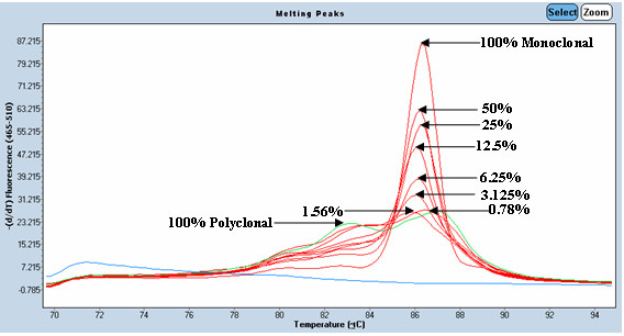
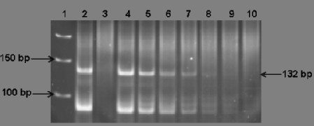
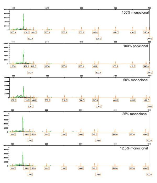
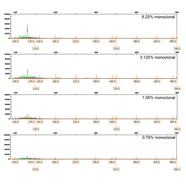
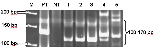
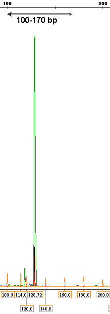
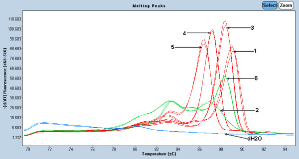
Introduction: Malignant lymphoma, especially non-Hodgkin lymphoma, is one of the most common hematologic malignancies in Thailand. The incidence rate as reported by Ministry of Public Health is 3.1 per 100,000 population in female whereas the rate in male is 4.5 per 100,000 population 1. At Siriraj Hospital, the new cases diagnosed as malignant lymphoma were 214.6 cases/year 2. The diagnosis of malignant lymphoma is often problematic, especially in early stages of the disease. Therefore, detection of antigen receptor gene rearrangement including T cell receptor (TCR) and immunoglobulin heavy chain (IgH) by polymerase chain reaction (PCR) assay has recently become a standard laboratory test for discrimination of reactive from malignant clonal lymphoproliferation 34. Analyzing DNA extracted from formalin-fixed, paraffin-embedded tissues by multiplex PCR techniques is more rapid, accurate and highly sensitive. Measuring the size of the amplicon from PCR analysis could be used to diagnose malignant lymphoma with monoclonal pattern showing specific and distinct bands detected on acrylamide gel electrophoresis. However, this technique has some limitations and some patients might require a further confirmation test such as GeneScan or fragment analysis 56.GeneScan technique or fragment analysis reflects size and peak of DNA by using capillary gel electrophoresis. This technique is highly sensitive and can detect 0.5-1% of clonal lymphoid cells. It measures the amplicons by using various fluorescently labeled primers at forward or reverse sides and a specific size standard. Using a Genetic Analyzer machine and GeneMapper software (Applied Bioscience, USA), the monoclonal pattern revealed one single, sharp and high peak at the specific size corresponding to acrylamide gel pattern, whereas the polyclonal pattern showed multiple and small peak condensed at the same size standard. This technique is the most sensitive and accurate technique; however, it usually requires high technical experience and is also of high cost 7. Therefore, rapid and more cost effective technique are being sought.LightCycler PCR performs the diagnostic detection of amplicon via melting curve analysis within 2 hours with the use of a specific dye 89. This dye consists of two types: one known as SYBR-Green I which is non specific and the other named as High Resolution Melting analysis (HRM) which is highly sensitive, more accurate and stable. Several reports demonstrated that this new instrument combined with DNA intercalating dyes can be used to discriminate sequence changes in PCR amplicon without manual handling of PCR product 1011. Therefore, current investigations using melting curve analysis are being developed 1213.In this study, three different techniques were compared to evaluate the suitability of LightCycler PCR with HRM as the clonal diagnostic tool for IgH gene rearrangement in B-cell non-Hogdkin lymphoma, i.e. thermocycler PCR followed by heteroduplex analysis and PAGE, GeneScan analysis and LightCycler PCR with HRM.
Figures
References
-
- Wiangnon S. Chapter II-20. Cancer in Thailand. 1998-2000. Vol. 4. Bangkok Medical Publisher. Bangkok. Thailand; 2007. Lymphoma; pp. 66–67.
-
- Trainor K, Brisco M, Wan J, Neoh S, Grist S, Morley A. Gene rearrangement in B and T lymphoproliferative disease detected by polymerase chain reaction. Blood. 1991;78:192. - PubMed
-
- Tai YC, Peh SC. Feasibility of T cell receptorγ (TCR γ) gene rearrangement on formalin fixed paraffin embedded tissues by PCR assays. Singapore Med J. 2003;44:250–255. - PubMed
-
- Assaf C, Hummel M, Dippel E, Goerdt S, Muller HH, Anagnostopoulos I, Orfanos CE, Stein H. High detection rate of T cell receptor beta chain rearrangements in T cell lymphoproliferations by family specific polymerase chain reaction in combination with the GeneScan technique and DNA sequencing. Blood. 2000;96:640–646. - PubMed
Publication types
MeSH terms
Substances
LinkOut - more resources
Full Text Sources
