

Effects of IL-6 and AG490 on regulation of Stat3 signaling pathway and invasion of human pancreatic cancer cells in vitro
- PMID: 20482858
- PMCID: PMC2883975
- DOI: 10.1186/1756-9966-29-51
Effects of IL-6 and AG490 on regulation of Stat3 signaling pathway and invasion of human pancreatic cancer cells in vitro
Abstract
Background: Signal transducer and activator of transcription 3 (Stat3) is a member of the Janus-activated kinase(Jak)/Stat signaling pathway. Abnormal activation of Stat3 plays a critical role in metastasis and invasion in varieties of human tumors including pancreatic cancer. This study aimed to investigate the mechanisms of activation and blocking of the Stat3 signaling pathway and its effects on invasion and metastasis of human pancreatic cancer cells.
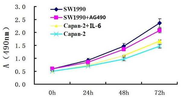
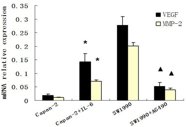
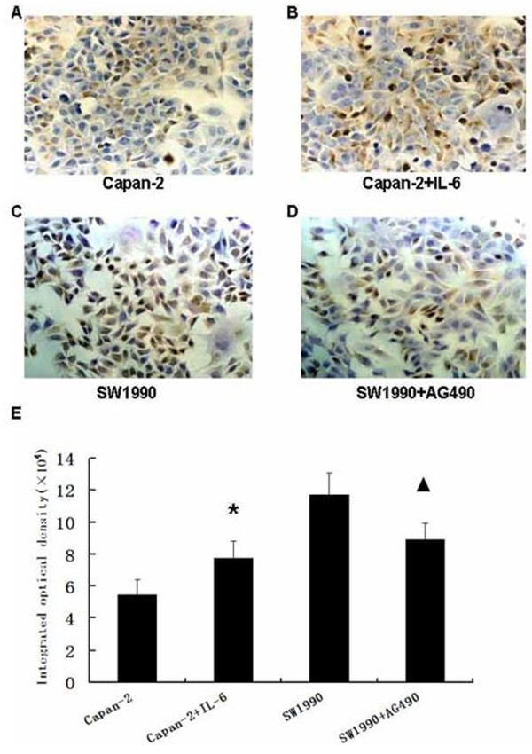
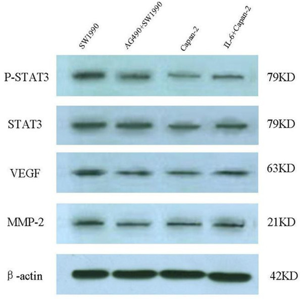
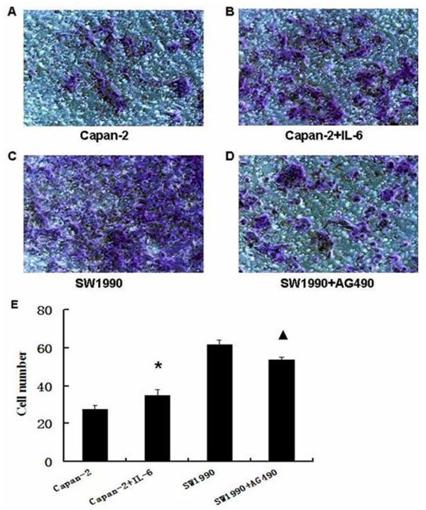
Methods: The Jak inhibitor AG490 and interleukin-6 (IL-6) were added to the culture media of human pancreatic cancer cells SW1990 and Capan-2, respectively. Cell growth was measured by MTT assays. Western blotting and immunocytochemistry were performed to detect phosphorylated Stat3 (p-Stat3) protein, while VEGF and MMP-2 mRNA and protein expression were examined with fluorescence quantitative polymerase chain reaction and Western blotting, respectively. The invasion ability of SW1990 and Capan-2 cells was determined by cell invasion assay.
Results: Stat3 was activated by IL-6 in Capan-2 cells; protein expression of p-Stat3 was increased significantly in Capan-2 cells. IL-6 remarkably promoted the growth of Capan-2 cells (P < 0.05), and VEGF and MMP-2 mRNA and protein expression were increased significantly. Also, IL-6 increased the invasion ability of Capan-2 cells. AG490 inhibited Stat3 activation in SW1990 cells. Western blotting and immunocytochemistry analysis showed that p-Stat3 protein expression was decreased significantly with AG490 treatment in SW1990 cells. AG490 remarkably inhibited the growth of Capan-2 cells (P < 0.05), and VEGF and MMP-2 mRNA and protein expression was decreased significantly. And AG490 decreased the invasion ability of SW1990 cells.
Conclusions: Abnormal activation of Stat3 plays an important role in the invasion and metastasis of pancreatic cancer. Activation and blocking of the Stat3 signaling pathway can affect invasion ability and expression of the VEGF and MMP-2 genes in pancreatic cancer cells. The Stat3 signaling pathway may provide a novel therapeutic target for treatment of pancreatic cancer.
Figures
Similar articles
-
[Inhibitory effect of AG490 on invasion and metastasis of human pancreatic cancer cells in vitro].Zhonghua Zhong Liu Za Zhi. 2006 Dec;28(12):890-3. Zhonghua Zhong Liu Za Zhi. 2006. PMID: 17533737 Chinese.
-
Inhibition of STAT3 activity with AG490 decreases the invasion of human pancreatic cancer cells in vitro.Cancer Sci. 2006 Dec;97(12):1417-23. doi: 10.1111/j.1349-7006.2006.00340.x. Epub 2006 Oct 19. Cancer Sci. 2006. PMID: 17054436 Free PMC article.
-
The effects and mechanisms of blockage of STAT3 signaling pathway on IL-6 inducing EMT in human pancreatic cancer cells in vitro.Neoplasma. 2011;58(5):396-405. doi: 10.4149/neo_2011_05_396. Neoplasma. 2011. PMID: 21744993
-
STAT3 signaling in pancreatic ductal adenocarcinoma: a candidate therapeutic target.Pathol Res Pract. 2023 May;245:154425. doi: 10.1016/j.prp.2023.154425. Epub 2023 Mar 24. Pathol Res Pract. 2023. PMID: 37019018 Review.
-
Targeting the IL-6/JAK/STAT3 signalling axis in cancer.Nat Rev Clin Oncol. 2018 Apr;15(4):234-248. doi: 10.1038/nrclinonc.2018.8. Epub 2018 Feb 6. Nat Rev Clin Oncol. 2018. PMID: 29405201 Free PMC article. Review.
Cited by
-
Focal adhesion kinase (FAK) activation by estrogens involves GPER in triple-negative breast cancer cells.J Exp Clin Cancer Res. 2019 Feb 6;38(1):58. doi: 10.1186/s13046-019-1056-8. J Exp Clin Cancer Res. 2019. PMID: 30728047 Free PMC article.
-
Plumbagin, a plant derived natural agent inhibits the growth of pancreatic cancer cells in in vitro and in vivo via targeting EGFR, Stat3 and NF-κB signaling pathways.Int J Cancer. 2012 Nov 1;131(9):2175-86. doi: 10.1002/ijc.27478. Epub 2012 Mar 20. Int J Cancer. 2012. PMID: 22322442 Free PMC article.
-
Significance of CXCR4, phosphorylated STAT3 and VEGF-A expression in resected non-small cell lung cancer.Exp Ther Med. 2011 May;2(3):517-522. doi: 10.3892/etm.2011.235. Epub 2011 Mar 21. Exp Ther Med. 2011. PMID: 22977534 Free PMC article.
-
The anti-dysenteric drug fraxetin enhances anti-tumor efficacy of gemcitabine and suppresses pancreatic cancer development by antagonizing STAT3 activation.Aging (Albany NY). 2021 Jul 28;13(14):18545-18563. doi: 10.18632/aging.203301. Epub 2021 Jul 28. Aging (Albany NY). 2021. PMID: 34320467 Free PMC article.
-
CEP55 Promotes Cell Motility via JAK2⁻STAT3⁻MMPs Cascade in Hepatocellular Carcinoma.Cells. 2018 Aug 8;7(8):99. doi: 10.3390/cells7080099. Cells. 2018. PMID: 30096813 Free PMC article.
References
Publication types
MeSH terms
Substances
LinkOut - more resources
Full Text Sources
Other Literature Sources
Medical
Miscellaneous
