. 2010 May 19:10:145.
doi: 10.1186/1471-2180-10-145.

Ultrastructure and molecular phylogenetic position of a novel euglenozoan with extrusive episymbiotic bacteria: Bihospites bacati n. gen. et sp. (Symbiontida)

Affiliations

Ultrastructure and molecular phylogenetic position of a novel euglenozoan with extrusive episymbiotic bacteria: Bihospites bacati n. gen. et sp. (Symbiontida)

Susana A Breglia et al. BMC Microbiol. .

Abstract

Background: Poorly understood but highly diverse microbial communities exist within anoxic and oxygen-depleted marine sediments. These communities often harbour single-celled eukaryotes that form symbiotic associations with different prokaryotes. During low tides in South-western British Columbia, Canada, vast areas of marine sand become exposed, forming tidal pools. Oxygen-depleted sediments within these pools are distinctively black at only 2-3 cm depth; these layers contain a rich variety of microorganisms, many of which are undescribed. We discovered and characterized a novel (uncultivated) lineage of heterotrophic euglenozoan within these environments using light microscopy, scanning and transmission electron microscopy, serial sectioning and ultrastructural reconstruction, and molecular phylogenetic analyses of small subunit rDNA sequences.

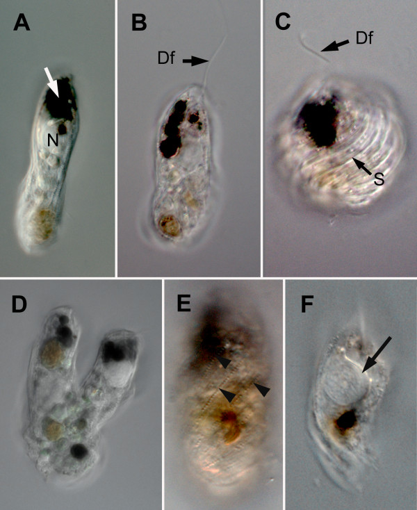
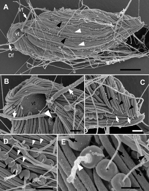
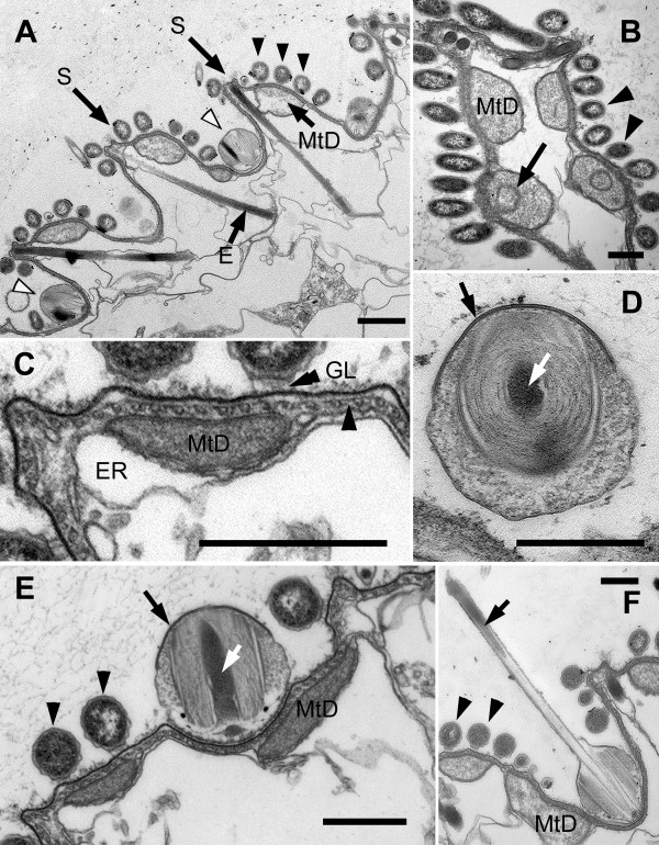
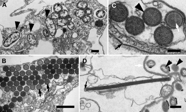
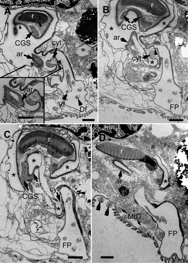
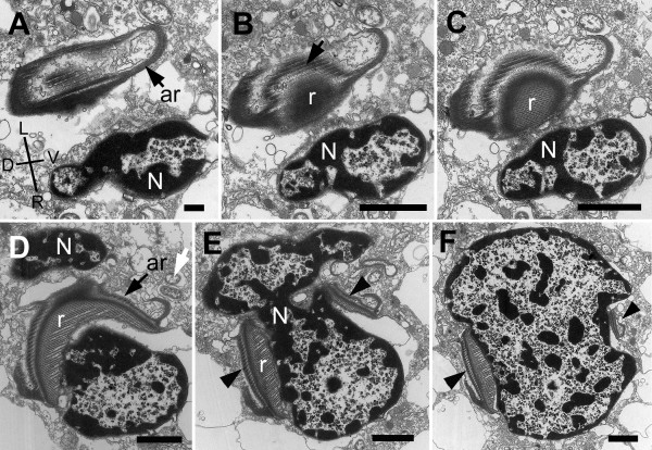
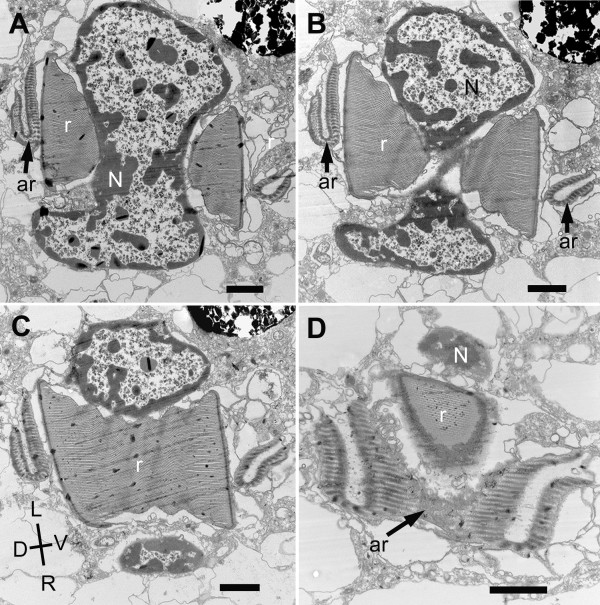
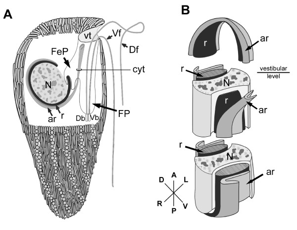
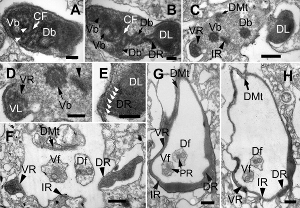
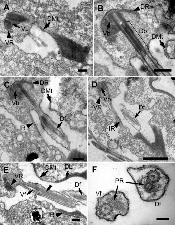
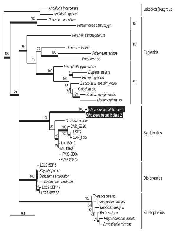
Results: Bihospites bacati n. gen. et sp. is a biflagellated microbial eukaryote that lives within low-oxygen intertidal sands and dies within a few hours of exposure to atmospheric oxygen. The cells are enveloped by two different prokaryotic episymbionts: (1) rod-shaped bacteria and (2) longitudinal strings of spherical bacteria, capable of ejecting an internal, tightly wound thread. Ultrastructural data showed that B. bacati possesses all of the euglenozoan synapomorphies. Moreover, phylogenetic analyses of SSU rDNA sequences demonstrated that B. bacati groups strongly with the Symbiontida: a newly established subclade within the Euglenozoa that includes Calkinsia aureus and other unidentified organisms living in low-oxygen sediments. B. bacati also possessed novel features, such as a compact C-shaped rod apparatus encircling the nucleus, a cytostomal funnel and a distinctive cell surface organization reminiscent of the pellicle strips in phagotrophic euglenids.

Conclusions: We characterized the ultrastructure and molecular phylogenetic position of B. bacati n. gen. et sp. Molecular phylogenetic analyses demonstrated that this species belongs to the Euglenozoa and currently branches as the earliest diverging member of the Symbiontida. This is concordant with ultrastructural features of B. bacati that are intermediate between C. aureus and phagotrophic euglenids, indicating that the most recent ancestor of the Symbiontida descended from phagotrophic euglenids. Additionally, the extrusive episymbionts in B. bacati are strikingly similar to so-called "epixenosomes", prokaryotes previously described in a ciliate species and identified as members of the Verrucomicrobia. These parallel symbioses increase the comparative context for understanding the origin(s) of extrusive organelles in eukaryotes and underscores how little we know about the symbiotic communities of marine benthic environments.


Figures

Figure 1
Figure 1
Light micrographs (LM) of living cells of Bihospites bacati n. gen. et sp. A. LM showing distinctive black bodies (white arrow) and the prominent nucleus (N) positioned near the anterior end of the cell. B. LM showing the extended dorsal flagellum (Df) that is inserted subapically. C. LM showing the dorsal flagellum (Df) and a contracted cell with raised helically arranged striations (S) on the surface. D. LM showing a cell dividing along the anteroposterior axis. E. LM showing rows of spherical-shaped bacterial episymbionts on the cell surface (arrowheads). F. LM showing the nucleus with a distinct thickening (arrow), providing evidence for the shape and orientation of the C-shaped rod apparatus.
Figure 2
Figure 2
Scanning electron micrographs (SEM) of Bihospites bacati n. gen. et sp. A. Ventral view of B. bacati showing a cell covered with rod-shaped and spherical-shaped episymbiotic bacteria (white arrowheads and black arrowheads, respectively), the vestibulum (vt), dorsal flagellum (Df) and ventral flagellum (Vf) (bar = 15 μm). B. High magnification of the vestibular opening (vt), showing the open cytostome (white arrowhead), and the dorsal (Df) and ventral flagella (Vf) without flagellar hairs. C. High magnification SEM showing the posterior end of B. bacati, in ventral view, and the external appearance of the raised articulation zones between S-shaped folds in the host cell surface (black arrowheads). The white arrows show pores on the cell surface. D. High magnification SEM showing the rod-shaped (white arrowheads) and spherical-shaped episymbionts. E. High magnification SEM of the spherical-shaped episymbionts showing discharged threads (black arrows) through an apical pore (bar = 0.5 μm). The white arrow shows the initial stages of the ejection process. (B-D bar = 1 μm).
Figure 3
Figure 3
Transmission electron micrographs (TEM) of the cell surface of Bihospites bacati n. gen. et sp. A. Cross-section of cell showing a series of S-shaped folds in the cell surface. Elongated extrusomes (E) positioned beneath the raised articulation zones between the S-shaped folds (S). Cell surface covered with rod-shaped bacteria (black arrowheads), in cross section, and spherical-shaped bacteria (white arrowheads). Mitochondrion-derived organelles (MtD) underlie the cell surface. (bar = 1 μm). B. TEM showing mitochondrion-derived organelles (MtD) with zero to two cristae (arrow). Arrowheads show transverse profiles of rod-shaped episymbionts on cell surface. C. High magnification TEM of the host cell surface showing glycogalyx (GL) connecting episymbionts to plasma membrane. Plasma membrane subtended by a thick layer of glycoprotein (double arrowhead) and a continuous row of microtubules linked by short 'arms' (arrowhead). Mitochondrion-derived organelles (MtD) positioned between the row of microtubules and the endoplasmic reticulum (ER). D. Oblique TEM section of spherical-shaped episymbiont showing electron-dense apical operculum (black arrow) and the extrusive thread coiled around a densely stained core region (white arrow). E. High magnification TEM of cell surface showing mitochondrion-derived organelles (MtD), rod-shaped episymbionts (arrowheads), and spherical-shaped episymbiont (black arrow) sitting within a corresponding concavity in the host cell. Core region of the spherical-shaped episymbiont (white arrow) in longitudinal section. F. TEM of spherical-shaped episymbiont showing discharged extrusive thread (arrow). Electron-dense material corresponding to the core is positioned at the tip of the discharged thread (arrow). Arrowheads indicate rod-shaped bacteria on cell surface (B-F bar = 500 nm).
Figure 4
Figure 4
Transmission electron micrographs (TEM) of Bihospites bacati n. gen. et sp. showing intracellular bacteria and extrusomes. A. TEM showing a cell containing numerous intracellular bacteria (arrowheads) within vacuoles. B. Transverse TEM showing a battery of extrusomes (arrows) (A, B, bar = 500 nm). C. High magnification TEM of extrusomes showing a dense outer region (arrowhead) and a granular core containing a lighter cruciform structure (white arrow). Black arrow denotes the plasma membrane of the host (bar = 100 nm). D. TEM showing a longitudinal section of an extrusome; the proximal end is indicated with a black arrow. Arrowheads denote rod-shaped bacteria on the cell surface (bar = 500 nm).
Figure 5
Figure 5
Transmission electron micrographs (TEM) of non-consecutive serial sections of Bihospites bacati n. gen. et sp. through the vestibular region of the cell. A. TEM showing the nucleus (N) with condensed chromatin, the dorsal side of the C-shaped rod apparatus consisting of the main rod (r) and the accessory rod (ar), and the vestibulum (vt). Several rod-shaped bacteria (black arrows) and spherical-shaped bacteria line inner surface of the vestibulum (vt) (bar = 10 μm). B. High magnification view of the C-shaped rod apparatus in Figure A showing the single row of microtubules (arrowheads) positioned at the junction between the tightly connected rod and accessory rod. Granular bodies (arrows) are present between the parallel lamellae that form the main rod (bar = 500 nm). C, D. Transverse TEMs showing the cytostomal funnel (cyt) and two separate lobes of the feeding pocket (arrowheads). Bacterial profiles can be seen inside the feeding pocket (arrows). Figure D uses color to distinguish between the feeding pocket (red), the vestibulum (blue), and the two branches of the flagellar pocket (green). E, F. Transverse TEMs at a more posterior level than in Figure C-D showing the posterior end of the main C-shaped rod (arrow) emerging within the posterior end of the feeding pocket. The cytostomal funnel (arrowheads) opens and fuses with the feeding pocket. Figure F uses color to distinguish between the feeding pocket (red), the vestibulum (blue), and the two branches of the flagellar pocket (green). (C-F bar = 2 μm).
Figure 6
Figure 6
Transmission electron micrographs (TEM) of non-consecutive serial sections through the flagellar apparatus and feeding pockets of Bihospites bacati n. gen. et sp. TEMs taken at levels posterior to those shown in Figure 5 and presented from anterior (A) to posterior (D). A. TEM showing the posterior end of the main C-shaped rod (r) embedded in an amorphous matrix (double arrowhead) and surrounded by a thick membrane with fuzzy material (arrowhead). At this level, the rod is associated with 'congregated globular structure' (CGS), and the striated fibres that form the accessory rod (ar) appear near the cytostomal funnel (cyt) at the junction between the feeding pocket and the flagellar pocket. Inset: TEM showing the accessory rod (ar) in a subsequent posterior section, as it starts to open up. Vf = ventral flagellum; Df = dorsal lagellum. B. TEM showing the separation (arrowhead) of the feeding pocket (asterisks) from the flagellar pocket (FP) near cytostomal funnel (cyt) and the expanding accessory rod (ar). C. TEM showing the diminishing feeding pocket (asterisks), the cytostomal funnel (cyt), and the separate flagellar pocket (FP). D. TEM showing the accessory rod (ar) with its characteristically folded shape becoming more tightly linked to the main rod (r). Lobes of the feeding pocket (asterisk) and the flagellar pocket (FP) are also still visible. MtD = mitochondrion-derived organelle; double arrowheads = spherical-shaped episymbionts. (bars = 2 μm).
Figure 7
Figure 7
Transmission electron micrographs (TEM) of non-consecutive serial sections through the anterior part of the nucleus of Bihospites bacati n. gen. et sp. Figures 7A-F are presented from anterior to posterior. A. TEM showing the nucleus (N) and the accessory rod (ar) surrounded by electron-dense material (Images are viewed from the anterior side of the cell: D, dorsal; L, left side of the cell; R, right side of the cell; V, ventral). B-C. TEMs showing the main rod (r) near the striated fibres (SF) of the accessory rod (arrow). D. TEM showing the left side of the nucleus (N) appearing behind the rod (r) and accessory rod (ar). The white arrow shows the presence of bacteria near the rod. E. TEMs showing the main rod (r) and the accessory rod (arrowheads) on the dorsal and ventral sides of the nucleus. F. Transverse TEM at the level of the vestibulum showing the disappearance of the ventral side of the main rod (r) and the drastic reduction of the accessory rod (arrowhead). Note the indentations in the nucleus for accommodating the main rod and accessory rod (A bar = 500 nm; B-F bar = 2 μm).
Figure 8
Figure 8
Transmission electron micrographs (TEM) of non-consecutive serial sections through the posterior part of the nucleus of Bihospites bacati n. gen. et sp. Figures 8A-D are presented from anterior to posterior. A-C. TEMs showing the rod (r) and the folded accessory rod (ar) nestled within indentations in the dorsal and ventral sides of the nucleus. The ventral part of the accessory rod runs close to the main rod for most of its length and extends toward the flagella on the ventral side of the cell. N = nucleus; D, dorsal; L, left side of the cell; R, right side of the cell; V, ventral; Images are viewed from the anterior side of the cell. D. TEMs showing the main rod (r) and the accessory rod (ar) reaching the posterior end of the nucleus (N). The main rod consists of parallel-arranged lamellae. Most of the nucleus and the main rod have disappeared from the section. The accessory rod (ar) consists of striated fibres that wrap around the main rod and the nucleus (bars = 2 μm).
Figure 9
Figure 9
Diagrams showing a reconstruction of the ultrastructure of Bihospites bacati n. gen. et sp. Relationships between C-shaped rod apparatus, nucleus, cytostomal funnel, feeding pocket, flagellar pocket and vestibulum, as inferred from serial transmission electron microscopy (TEM), scanning electron microscopy (SEM), and light microscopy (LM). A. Cell viewed from the right side showing the positions of the nucleus (N), the C-shaped main rod (r), the accessory rod (ar), and the cytostomal funnel (cyt) in relation to the feeding pocket (FeP), the flagellar pocket (FP) and the vestibulum (vt); Vf = ventral flagellum; Df = dorsal flagellum; Db = dorsal basal body; Vb = ventral basal body. B. Diagram emphasizing the relationship between nucleus (N), main rod (r), and folded accessory rod (ar). The diagram is divided into three sections; and the nucleus removed from the top section for clarity. Posterior end of the main rod positioned at the level of the vestibulum on the ventral side of the nucleus. This rod extends posteriorly and then encircles the posterior, dorsal and anterior ends of the nucleus before terminating on the ventral side of the nucleus just above the vestibulum; therefore, this rod is C-shaped. The folded accessory rod runs along the C-shaped main rod for most of its length, terminating at the same point just above the vestibulum; however, on the ventral side of the nucleus, the posterior end of the accessory rod extends both anteriorly, defining the cytostomal funnel (cyt), and ventrally toward the ventral basal body.
Figure 10
Figure 10
TEM micrographs showing sections of basal bodies, flagellar roots and associated structures, of Bihospites bacati n. gen. et sp. A-H from proximal to distal end of flagellar pocket. A-C. Non-consecutive serial sections showing origin and organization of flagellar pocket. A. High magnification TEM of proximal region of basal bodies showing dorsal and ventral basal bodies (Db and Vb) linked by a connecting fibre (CF). Basal bodies with cartwheel structures associated to electron-dense fibres (arrowheads). B. TEM showing accessory dorsal and ventral basal bodies (Db' and Vb') on the left of the two main basal bodies. Dorsal root (DR) connects to electron-dense body (dorsal lamella=DL), on right side of Db. C. TEM showing intermediate root (IR) associated with right side of Vb. Ventral root (VR) associated with electron-dense material that becomes ventral lamella (VL). Row of dorsal microtubules (DMt), not associated with basal bodies. D. Detail of ventral side of Figure C showing Vb, VR formed by four microtubules, VL and intermediate root (arrowhead), initially composed of eight microtubules. E. Detail of dorsal side of Figure C showing DR, with six microtubules (white arrowheads), and DL. F. TEM showing three flagellar roots and DMt around flagellar pocket. Df = dorsal flagellum; Vf = ventral flagellum. G-H. Non-consecutive serial TEM sections of flagellar pocket showing Df and Vf with paraxial rods (PR), flagellar roots, DMt of microtubules lining flagellar pocket, and DL and VL. (A-B and D-E bars = 200 nm; C and F bars = 500 nm; G-H bars = 2 μm)
Figure 11
Figure 11
Transmission electron micrographs (TEM) of Bihospites bacati n. gen. et sp. showing the emergence and organization of the flagella. A. Longitudinal TEM through the electron-dense region near the origin of the basal bodies. The ventral root (VR) originates from the ventral basal body (Vb). A row of microtubules (DMt) lines the dorsal side of the incipient flagellar pocket. B. Longitudinal TEM through the dorsal flagellum showing the dorsal basal body (Db) associated with the dorsal flagellar root (DR), the ventral basal body (Vb), and the dorsal microtubules (DMt). C-D. TEM sections showing the dorsal flagellum (Df) and the intermediate root (IR) associated with the ventral basal body (Vb). E. TEM showing oblique sections through both flagella and the positions of the VR, IR and DMt in the flagellar pocket. The electron-dense material from which the flagellar apparatus originated in Figure A elongates to form the dorsal lamella (DL). The double arrowheads show the paraxial rod in the ventral flagellum (Vf). F. Transverse TEM of the Df and Vf showing the 9+2 arrangement of microtubules in the axoneme and the heteromorphic paraxial rods (PR). (A-E bars = 500 nm; F bar = 200 nm)
Figure 12
Figure 12
Phylogenetic position of Bihospites bacati n. gen. et sp. within the Euglenozoa as inferred from small subunit (SSU) rDNA sequences. Maximum likelihood (ML) analysis of 35 euglenozoan taxa, rooted with two jakobids (Andalucia incarcerata and A. godoyi). Only ML boostraps greater then 50% are shown. Thick branches correspond to Bayesian posterior probabilities over 0.95. Ba, bacterivorous taxa; Eu, eukaryovorous taxa; Ph, photosynthetic taxa.

Similar articles

Cited by

References

    1. Leander BS, Farmer MA. Comparative Morphology of the Euglenid Pellicle. I. Patterns of strips and pores. J Eukaryot Microbiol. 2000;47:469–479. doi: 10.1111/j.1550-7408.2000.tb00076.x. - DOI - PubMed
    1. Triemer RE, Farmer MA. In: The Biology of Free-living Heterotrophic Flagellates. Patterson DJ, Larsen J, editor. Clarendon Press, Oxford; 1991. The ultrastructural organization of the heterotrophic euglenids and its evolutionary implications; pp. 205–217.
    1. Leander BS, Triemer RE, Farmer MA. Character evolution in heterotrophic euglenids. Eur J Protistol. 2001;37:337–356. doi: 10.1078/0932-4739-00842. - DOI
    1. Simpson AGB, Lukes J, Roger AJ. The evolutionary history of kinetoplastids and their kinetoplasts. Mol Biol Evol. 2002;19:2071–2083. - PubMed
    1. Kivic PA, Walne PL. An evaluation of the possible phylogenetic relationship between euglenophyta and kinetoplastida. Orig Life. 1984;13:269–288. doi: 10.1007/BF00927177. - DOI

Publication types

MeSH terms

Associated data

LinkOut - more resources