. 2010;11(5):R55.
doi: 10.1186/gb-2010-11-5-r55. Epub 2010 May 19.

Studies on Xenopus laevis intestine reveal biological pathways underlying vertebrate gut adaptation from embryo to adult

Affiliations

Studies on Xenopus laevis intestine reveal biological pathways underlying vertebrate gut adaptation from embryo to adult

Rachel A Heimeier et al. Genome Biol. 2010.

Abstract

Background: To adapt to its changing dietary environment, the digestive tract is extensively remodeled from the embryo to the adult during vertebrate development. Xenopus laevis metamorphosis is an excellent model system for studying mammalian gastrointestinal development and is used to determine the genes and signaling programs essential for intestinal development and maturation.

Results: The metamorphosing intestine can be divided into four distinct developmental time points and these were analyzed with X. laevis microarrays. Due to the high level of conservation in developmental signaling programs and homology to mammalian genes, annotations and bioinformatics analysis were based on human orthologs. Clustering of the expression patterns revealed co-expressed genes involved in essential cell processes such as apoptosis and proliferation. The two largest clusters of genes have expression peaks and troughs at the climax of metamorphosis, respectively. Novel conserved gene ontology categories regulated during this period include transcriptional activity, signal transduction, and metabolic processes. Additionally, we identified larval/embryo- and adult-specific genes. Detailed analysis revealed 17 larval specific genes that may represent molecular markers for human colonic cancers, while many adult specific genes are associated with dietary enzymes.

Conclusions: This global developmental expression study provides the first detailed molecular description of intestinal remodeling and maturation during postembryonic development, which should help improve our understanding of intestinal organogenesis and human diseases. This study significantly contributes towards our understanding of the dynamics of molecular regulation during development and tissue renewal, which is important for future basic and clinical research and for medicinal applications.


Figures
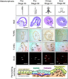
Figure 1
Figure 1
Morphological, histological and gene expression changes associated with X. laevis intestinal remodeling during natural metamorphosis. Representative metamorphic stages and the corresponding intestine evaluated with H&E (arrowheads indicate the islets of proliferating cells), TUNEL assay (arrows indicate the apoptotic cells), and BrdU immunohistochemistry (arrows indicate proliferating cells). Scale bar = 100 μm. AE:adult epithelial; Ct: connective tissue; Ep: epithelium; m: muscle; Ty: typhlosole. The schematic representation at the bottom summarizes the major changes associated with the stage-dependent transition.
Figure 2
Figure 2
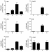
Expression changes of TRβ, THb/ZIP, ST3, XHH, GelA and IFABP, which are established intestinal remodeling markers, during natural development. The results are expressed relative to the control rpl8.
Figure 3
Figure 3
Genes significantly up- and down-regulated in the intestine during natural metamorphosis at specific stages when compared to stage 53. Venn diagrams showing the number of genes significantly (a) up-regulated and (b) down-regulated in the intestine during natural metamorphosis when the indicated stages were compared to stage 53 by microarray. (c) Temporal changes in gene expression during natural development visualized by heatmap. Normalized mean-centered expression levels for each gene are shown with black representing mean expression levels of four stages for a given gene, and green and red indicating lower or higher than the average as shown in the color legend.
Figure 4
Figure 4
Confirmation of gene regulation patterns identified by microarray with RT-qPCR. (a) Microarray. (b) RT-qPCR. GenBank accession numbers are shown above the graphs. The vertical axis in (a) shows the normalized log intensity of the expression and in (b) shows the expression of the genes with stage 53 arbitrarily set to 1.
Figure 5
Figure 5
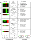
Regulated genes can be grouped into six clusters based on developmental regulation patterns. The number of genes in each cluster is indicated in the schematic diagram. (a, b) Clusters 1 and 2 represent genes that are predominantly regulated at metamorphic climax, with the former following the endogenous T3 concentration. (c, d) Clusters 3 and 4 include genes with higher levels of expression in tadpoles and frogs (larval- and adult-enriched genes), respectively. (e, f) Clusters 5 and 6 are genes up- or down-regulated mainly at stage 58. All clusters were evaluated by GO analysis and two or more examples of the significantly regulated GO categories that had >60 genes (clusters 1 and 2) and >5 genes (clusters 3 to 6) regulated during metamorphosis are listed. A complete list of GO categories associated with each cluster is listed in Table S6 in Additional file 1. PCA: principal component analysis.
Figure 6
Figure 6
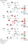
Temporal regulation of a significantly regulated biological pathway, the TGF-β pathway, during intestinal remodeling. Genes that are up- or down-regulated at stages 58, 61 and 66 relative to stage 53 are shown in red and green, respectively.
Figure 7
Figure 7
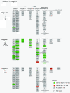
Temporal regulation of the electron transport pathway during intestinal remodeling. Genes that are up- or down-regulated at stages 58, 61 and 66 relative to stage 53 are shown in red and green, respectively.
Figure 8
Figure 8
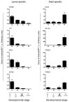
The expression of larval- and adult-specific genes during natural metamorphosis. Larval- and adult-specific genes analyzed by RT-qPCR, confirming the larval- and adult-specific designation based on microarray analysis.
Figure 9
Figure 9
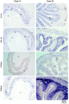
In situ hybridization of the four adult-specific genes with intestinal cross-sections at stages 53 and 66. Scale bar = 100 μm. AE: adult epithelium; Ct: connective tissue; Ep: epithelium; L: lumen; M: muscle; Ty: typhlosole.
Figure 10
Figure 10
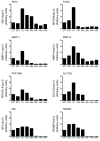
Gene expression during mouse development. RT-qPCR analysis was carried out for the indicated genes on mouse intestinal RNA at indicated stages. Note that the expression patterns of mouse EB13, FCNA, MMP11, MMP14, PPP1RA1 and SLC7A2 were similar to those of their homologs in Xenopus during metamorphosis as observed by microarray, that is, there were higher levels of expression shortly after birth in the mouse when T3 levels are high, just like at metamorphic climax. The expression of NPL and RENBP represent profiles of genes that differ from those in Xenopus. All RT-qPCR results are expressed relative to the control RPS13, with the expression of the genes at E17 arbitrarily set to 1.

Similar articles

Cited by

References

    1. Dauca M, Bouziges F, Colin S, Kedinger M, Keller MK, Schilt J, Simon-Assmann P, Haffen K. Development of the vertebrate small intestine and mechanisms of cell differentiation. Int J Dev Biol. 1990;34:205–218. - PubMed
    1. Cheng H, Leblond CP. Origin, differentiation and renewal of the four main epithelial cell types in the mouse small intestine. III. Entero-endocrine cells. Am J Anat. 1974;141:503–519. doi: 10.1002/aja.1001410405. - DOI - PubMed
    1. van der Flier LG, Clevers H. Stem cells, self-renewal, and differentiation in the intestinal epithelium. Annu Rev Physiol. 2009;71:241–260. doi: 10.1146/annurev.physiol.010908.163145. - DOI - PubMed
    1. Simon TC, Gordon JI. Intestinal epithelial cell differentiation: new insights from mice, flies and nematodes. Curr Opin Genet Dev. 1995;5:577–586. doi: 10.1016/0959-437X(95)80026-3. - DOI - PubMed
    1. Segal GH, Petras RE. In: Histology for Pathologists. Sternberg SS, editor. New York.: Raven Press, Ltd; 1992. Small intestine; pp. 547–571.

Publication types

MeSH terms

Substances