. 2010 May 19;98(10):2053-62.
doi: 10.1016/j.bpj.2009.12.4317.

Alzheimer's abeta(1-40) amyloid fibrils feature size-dependent mechanical properties

Affiliations

Alzheimer's abeta(1-40) amyloid fibrils feature size-dependent mechanical properties

Zhiping Xu et al. Biophys J. .

Abstract

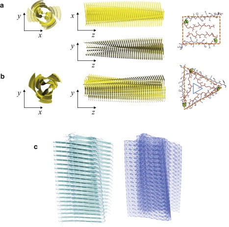
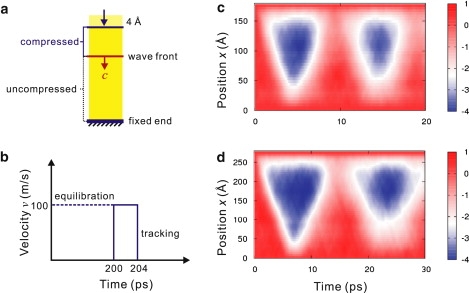
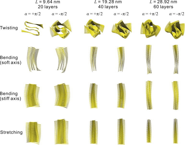
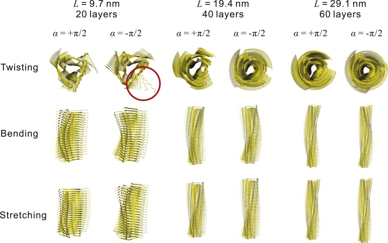
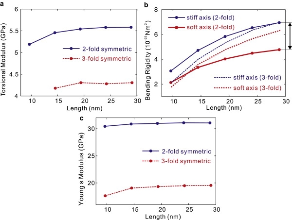
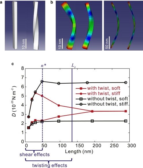
Amyloid fibrils are highly ordered protein aggregates that are associated with several pathological processes, including prion propagation and Alzheimer's disease. A key issue in amyloid science is the need to understand the mechanical properties of amyloid fibrils and fibers to quantify biomechanical interactions with surrounding tissues, and to identify mechanobiological mechanisms associated with changes of material properties as amyloid fibrils grow from nanoscale to microscale structures. Here we report a series of computational studies in which atomistic simulation, elastic network modeling, and finite element simulation are utilized to elucidate the mechanical properties of Alzheimer's Abeta(1-40) amyloid fibrils as a function of the length of the protein filament for both twofold and threefold symmetric amyloid fibrils. We calculate the elastic constants associated with torsional, bending, and tensile deformation as a function of the size of the amyloid fibril, covering fibril lengths ranging from nanometers to micrometers. The resulting Young's moduli are found to be consistent with available experimental measurements obtained from long amyloid fibrils, and predicted to be in the range of 20-31 GPa. Our results show that Abeta(1-40) amyloid fibrils feature a remarkable structural stability and mechanical rigidity for fibrils longer than approximately 100 nm. However, local instabilities that emerge at the ends of short fibrils (on the order of tens of nanometers) reduce their stability and contribute to their disassociation under extreme mechanical or chemical conditions, suggesting that longer amyloid fibrils are more stable. Moreover, we find that amyloids with lengths shorter than the periodicity of their helical pitch, typically between 90 and 130 nm, feature significant size effects of their bending stiffness due the anisotropy in the fibril's cross section. At even smaller lengths (50 nm), shear effects dominate lateral deformation of amyloid fibrils, suggesting that simple Euler-Bernoulli beam models fail to describe the mechanics of amyloid fibrils appropriately. Our studies reveal the importance of size effects in elucidating the mechanical properties of amyloid fibrils. This issue is of great importance for comparing experimental and simulation results, and gaining a general understanding of the biological mechanisms underlying the growth of ectopic amyloid materials.


Figures

Figure 1
Figure 1
Structure of twofold (a) and threefold (b) symmetric assemblies of Aβ(1-40) amyloid fibrils. Both structures have an intrinsic twist along the fibril axis (10,36). In the twofold fibril, two β-strands align in parallel and form a close contact, whereas in the threefold fibril, a hydrophobic core nanopore is formed in the center of the triangular structure. The visualizations on the outermost right show the geometry of a single protofibril layer for each morphology. (c) Full-atomistic and corresponding ENM representations of a twofold symmetric amyloid fibril.
Figure 2
Figure 2
Compressive WFT approach carried out using full-atomistic simulations. (a) Compression load is applied by prescribing a constant velocity to the Cα atoms in the two top protofibrils layers. (b) Loading history, consisting of an equilibration process for 200 ps, a pulse compression process with constant velocity applied to two top protofibril layers, and a wave-tracking process. (c) Displacement of Cα atoms in the twofold symmetric fibrils (length L = 19.28 nm), resulting in a measured wave speed of 4000 m/s. The color describes the axial displacement of the Cα atoms as a function of time (x-axis) and their initial positions (y-axis). (d) Displacement of Cα atoms in the threefold symmetric fibrils (length L = 29.1 nm), resulting in a measured wave speed of 3860 m/s.
Figure 3
Figure 3
Lowest-order collective modes of twofold symmetric amyloid fibrils, including twisting, bending along different axes (soft and stiff), and stretching modes. For shorter fibrils, the soft and stiff axes are well defined, showing significant differences between the two bending modes (where the one with lower frequency corresponds to bending around the soft axis). We find that there is also a significant coupling within these modes, particularly in bending and stretching.
Figure 4
Figure 4
Lowest-order collective modes of threefold symmetric amyloid fibrils, including twisting, bending, and stretching modes. The threefold symmetric triangular symmetry of the cross section results in a degenerated transverse bending mode.
Figure 5
Figure 5
Elastic properties of amyloid fibrils as a function of length L, as obtained from ENM calculations. (a) Torsional modulus G. (b) Bending rigidity D = YI along the soft and stiff axes of two- and threefold symmetric amyloid fibrils (with smaller and larger moments of inertia, respectively). (c) Young's modulus Y. All parameters are shown as a function of the amyloid fibril length up to ≈30 nm.
Figure 6
Figure 6
Results of normal-mode analysis based on three-dimensional, finite-element simulations. (a) The geometry of untwisted and twisted amyloid fibrils (length L = 48.2 nm (100 layers)). (b) The shape of bending modes along soft and stiff axes, respectively (for fibril lengths L = 48.2 nm (100 layers) and L = 192.8 nm (400 layers)). The bending modes for longer fibrils illustrate the degeneracy of the soft and stiff modes. The color represents displacement amplitude, scaled up by a factor of 80 for visualization. (c) Bending rigidity D of twofold symmetric amyloid fibrils with (solid symbols) and without (open symbols) the intrinsic twist along the fibril axis. The length-dependent bending rigidity values show significant size effects resulting from two distinct mechanisms: 1), at low aspect ratios n < n = 10, the bending rigidities calculated based on the Euler-Bernoulli theory increase with fibril length because of the unaccounted-for contributions from rotational and shear effects (for details, see Supporting Material); and 2), there is a geometric effect from the intrinsic twist on the resulting bending properties. For the untwisted fibril, the bending rigidities associated with the soft and stiff modes converge to distinct constant values for larger L. For twisted fibrils, when L is larger than the helical pitch length Lp, the difference between the soft and stiff modes disappears and their bending rigidity approaches the same value.

Similar articles

Cited by

References

    1. Chiti F., Dobson C.M. Protein misfolding, functional amyloid, and human disease. Annu. Rev. Biochem. 2006;75:333–366. - PubMed
    1. Dobson C.M. An overview of protein misfolding diseases. In: Buchner J., Kiefhaber T., editors. Protein Folding Handbook. Wiley-VCH Verlag GMBH & Co. KgaA; Weinheim, Germany: 2005. pp. 1093–1113.
    1. Jiménez J.L., Nettleton E.J., Saibil H.R. The protofilament structure of insulin amyloid fibrils. Proc. Natl. Acad. Sci. USA. 2002;99:9196–9201. - PMC - PubMed
    1. Nelson R., Eisenberg D. Recent atomic models of amyloid fibril structure. Curr. Opin. Struct. Biol. 2006;16:260–265. - PubMed
    1. Harper J.D., Lieber C.M., Lansbury P.T. Atomic force microscopic imaging of seeded fibril formation and fibril branching by the Alzheimer's disease amyloid-β protein. Chem. Biol. 1997;4:951–959. - PubMed