

Structural hierarchy governs fibrin gel mechanics
- PMID: 20483337
- PMCID: PMC2872216
- DOI: 10.1016/j.bpj.2010.01.040
Structural hierarchy governs fibrin gel mechanics
Abstract
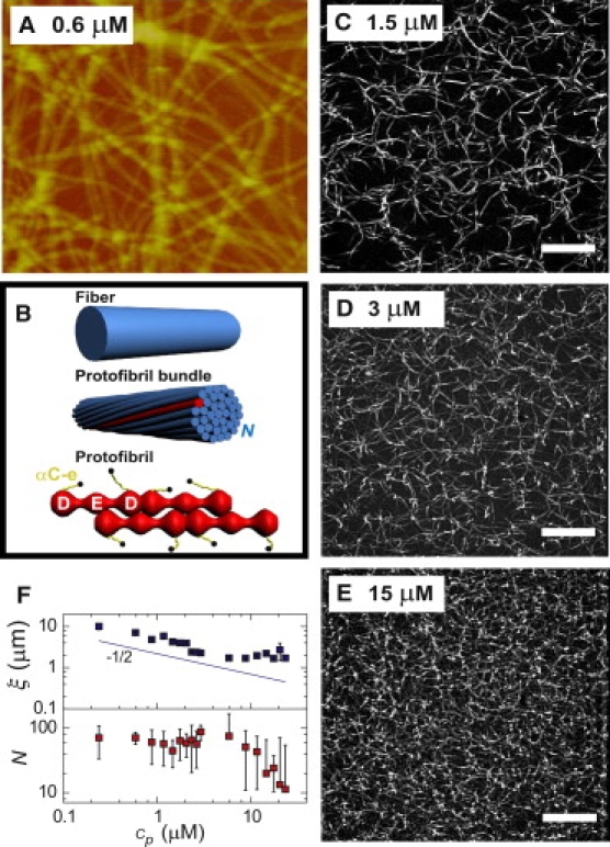
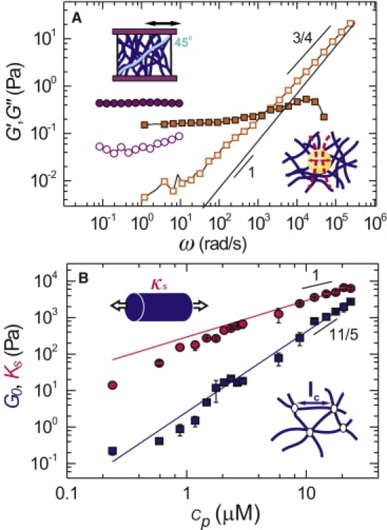
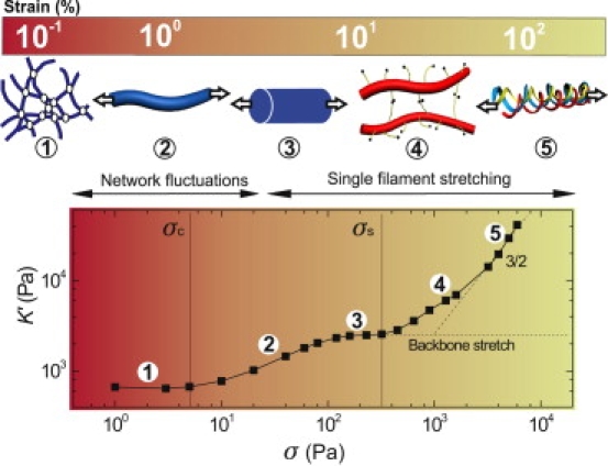
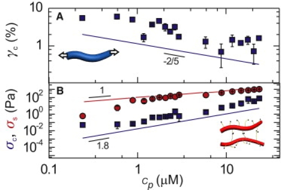
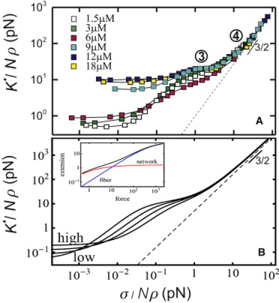
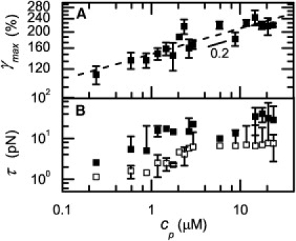
Fibrin gels are responsible for the mechanical strength of blood clots, which are among the most resilient protein materials in nature. Here we investigate the physical origin of this mechanical behavior by performing rheology measurements on reconstituted fibrin gels. We find that increasing levels of shear strain induce a succession of distinct elastic responses that reflect stretching processes on different length scales. We present a theoretical model that explains these observations in terms of the unique hierarchical architecture of the fibers. The fibers are bundles of semiflexible protofibrils that are loosely connected by flexible linker chains. This architecture makes the fibers 100-fold more flexible to bending than anticipated based on their large diameter. Moreover, in contrast with other biopolymers, fibrin fibers intrinsically stiffen when stretched. The resulting hierarchy of elastic regimes explains the incredible resilience of fibrin clots against large deformations.
Copyright 2010 Biophysical Society. Published by Elsevier Inc. All rights reserved.
Figures
Similar articles
-
Multi-scale strain-stiffening of semiflexible bundle networks.Soft Matter. 2016 Feb 21;12(7):2145-56. doi: 10.1039/c5sm01992c. Epub 2016 Jan 13. Soft Matter. 2016. PMID: 26761718
-
Micro-tensile rheology of fibrous gels quantifies strain-dependent anisotropy.Acta Biomater. 2024 Jun;181:272-281. doi: 10.1016/j.actbio.2024.03.028. Epub 2024 Apr 28. Acta Biomater. 2024. PMID: 38685460
-
Nonlinear elasticity of stiff filament networks: strain stiffening, negative normal stress, and filament alignment in fibrin gels.J Phys Chem B. 2009 Mar 26;113(12):3799-805. doi: 10.1021/jp807749f. J Phys Chem B. 2009. PMID: 19243107 Free PMC article.
-
Fibrin mechanical properties and their structural origins.Matrix Biol. 2017 Jul;60-61:110-123. doi: 10.1016/j.matbio.2016.08.003. Epub 2016 Aug 20. Matrix Biol. 2017. PMID: 27553509 Free PMC article. Review.
-
Fibrin gels and their clinical and bioengineering applications.J R Soc Interface. 2009 Jan 6;6(30):1-10. doi: 10.1098/rsif.2008.0327. J R Soc Interface. 2009. PMID: 18801715 Free PMC article. Review.
Cited by
-
Compressive instabilities enable cell-induced extreme densification patterns in the fibrous extracellular matrix: Discrete model predictions.PLoS Comput Biol. 2024 Jul 1;20(7):e1012238. doi: 10.1371/journal.pcbi.1012238. eCollection 2024 Jul. PLoS Comput Biol. 2024. PMID: 38950077 Free PMC article.
-
Spatiotemporal control of micromechanics and microstructure in acoustically-responsive scaffolds using acoustic droplet vaporization.Soft Matter. 2020 Jul 22;16(28):6501-6513. doi: 10.1039/d0sm00753f. Soft Matter. 2020. PMID: 32597450 Free PMC article.
-
Cell-matrix reciprocity in 3D culture models with nonlinear elasticity.Bioact Mater. 2021 Aug 14;9:316-331. doi: 10.1016/j.bioactmat.2021.08.002. eCollection 2022 Mar. Bioact Mater. 2021. PMID: 34820573 Free PMC article. Review.
-
Buffers Strongly Modulate Fibrin Self-Assembly into Fibrous Networks.Langmuir. 2017 Jun 27;33(25):6342-6352. doi: 10.1021/acs.langmuir.7b00527. Epub 2017 Jun 13. Langmuir. 2017. PMID: 28558246 Free PMC article.
-
Biophysical Mechanisms Mediating Fibrin Fiber Lysis.Biomed Res Int. 2017;2017:2748340. doi: 10.1155/2017/2748340. Epub 2017 May 28. Biomed Res Int. 2017. PMID: 28630861 Free PMC article. Review.
References
-
- Laurens N., Koolwijk P., Maat M.D. Fibrin structure and wound healing. J. Thromb. Haemost. 2008;4:932–939. - PubMed
-
- Kroll M.H., Hellums J.D., Moake J.L. Platelets and shear stress. Blood. 1996;88:1525–1541. - PubMed
-
- Shah J., Janmey P. Strain hardening of fibrin gels and plasma clots. Rheol. Acta. 1997;36:262–268.
Publication types
MeSH terms
Substances
LinkOut - more resources
Full Text Sources
Other Literature Sources
