. 2010 May 19;98(10):2299-308.
doi: 10.1016/j.bpj.2010.01.056.

Amyloid-beta fibrillogenesis seeded by interface-induced peptide misfolding and self-assembly

Affiliations

Amyloid-beta fibrillogenesis seeded by interface-induced peptide misfolding and self-assembly

Eva Y Chi et al. Biophys J. .

Abstract

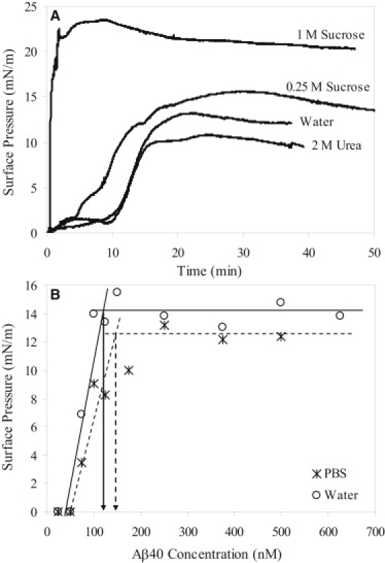
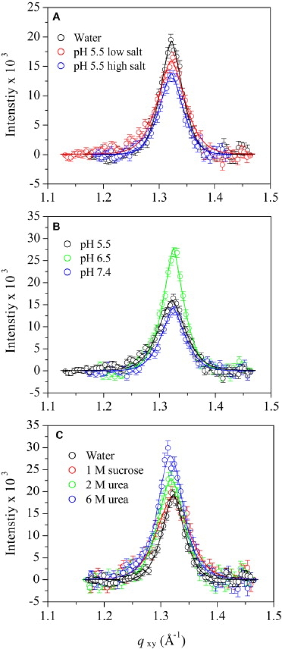
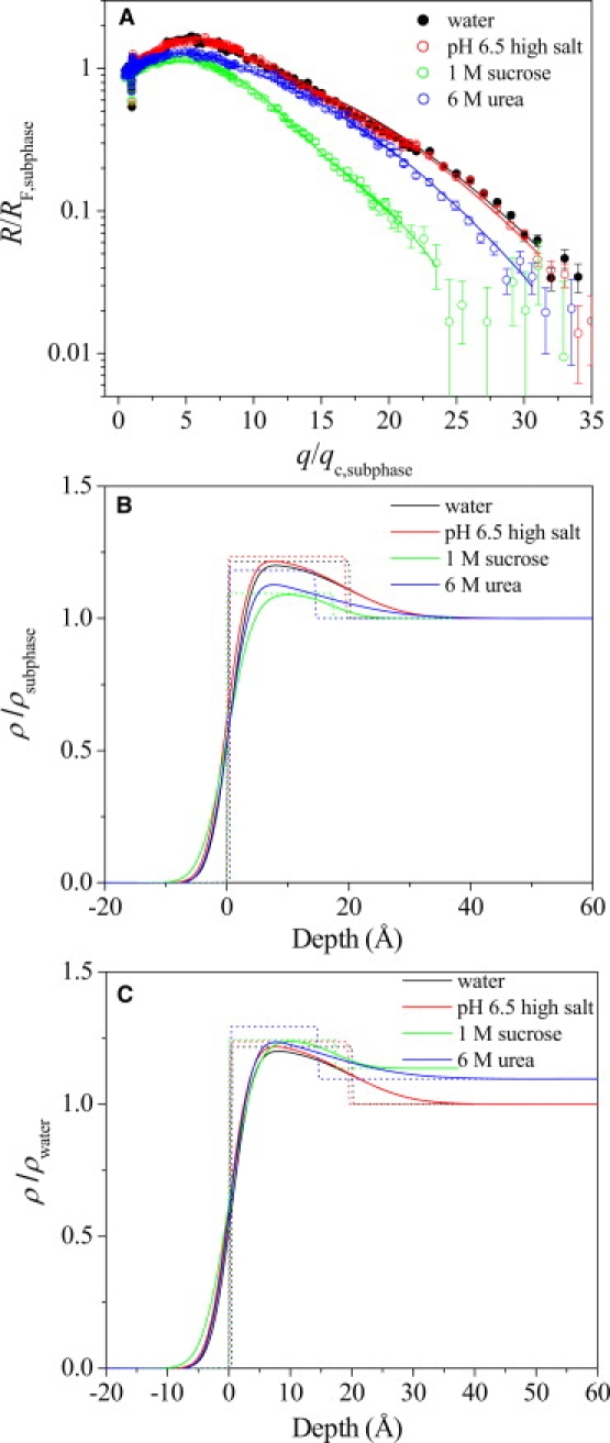
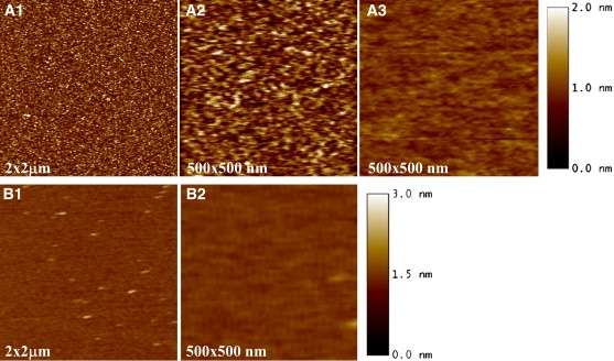
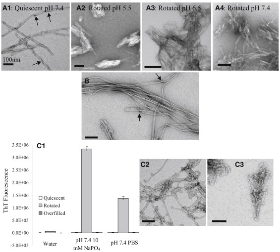
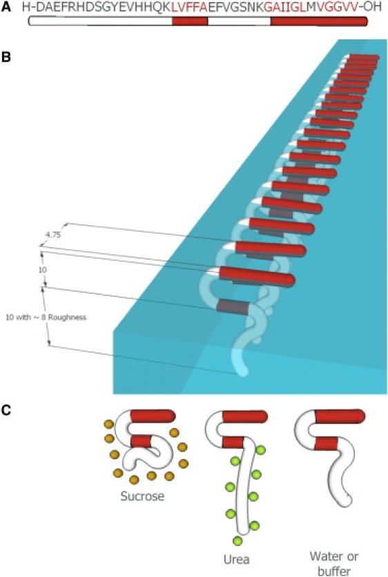
The amphipathicity of the natively unstructured amyloid-beta (Abeta40) peptide may play an important role in its aggregation into beta-sheet rich fibrils, which is linked to the pathogenesis of Alzheimer's disease. Using the air/subphase interface as a model interface, we characterized Abeta's surface activity and its conformation, assembly, and morphology at the interface. Abeta readily adsorbed to the air/subphase interface to form a 20 A thick film and showed a critical micelle concentration of approximately 120 nM. Abeta adsorbed at the air/subphase exhibited in-plane ordering that gave rise to Bragg peaks in grazing-incidence x-ray diffraction measurements. Analysis of the peaks showed that the air/subphase interface induced Abeta to fold into a beta-sheet conformation and to self-assemble into approximately 100 A-sized ordered clusters. The formation of these clusters at the air/subphase interface was not affected by pH, salts, or the presence of sucrose or urea, which are known to stabilize or denature native proteins, suggesting that interface-driven Abeta misfolding and assembly are strongly favored. Furthermore, Abeta at the interface seeded the growth of fibrils in the bulk with a distinct morphology compared to those formed by homogeneous nucleation. Our results indicate that interface-induced Abeta misfolding may serve as a heterogeneous, nucleation-controlled aggregation mechanism for Abeta fibrillogenesis in vivo.


Figures

Figure 1
Figure 1
(A) Adsorption isotherms of Aβ in various subphase conditions. (B) Determination of the CMC values of Aβ in water (circles and lines) and PBS (stars and dashed lines). The extrapolated CMC values for Aβ in water and PBS were ∼120 and 145 nM, respectively.
Figure 2
Figure 2
Background-subtracted Bragg peak profiles from GIXD data of Aβ adsorbed to the air/subphase interface at 30°C. (A) Effect of ionic strength on the Bragg peak profile of Aβ at pH 5.5. Low-salt buffer contained 10 mM sodium acetate, and high-salt buffer contained 10 mM sodium acetate and 140 mM NaCl. (B) Effect of pH on the Bragg peak profiles of Aβ in 10 mM salt buffers. (C) Effect of neutral cosolutes on the Bragg peak profiles of Aβ in water. Solid lines are fits to the data.
Figure 3
Figure 3
(A) Open symbols show the Fresnel normalized reflectivity data (R/RF) and fits for Aβ adsorbed to the air/subphase interface at 30°C. The solid lines represent the model-dependent fit to the reflectivity curves obtained from StochFit fitting routine. A one-box model with two roughness parameters was used to fit all of the reflectivity data (dashed lines). Corresponding electron density profiles of Aβ adsorbed at the air/subphase interface (B) normalized to ρsubphase and (C) normalized to ρwater are shown. Both smeared (solid line) and unsmeared (dotted line) electron density profiles are plotted. Negative depth values denote air, and positive depth values denote the subphase.
Figure 4
Figure 4
AFM images of Aβ transferred from the (A1 and A2) air/water and (B1and B2) air/PBS interfaces. (A3) Aβ adsorbed to a mica surface from the bulk was also imaged.
Figure 5
Figure 5
TEM images of 25 μM Aβ fibrils obtained in various solution conditions using different incubation methods: (A1) Aβ quiescently incubated in pH 7.4 10 mM sodium phosphate buffer for 36 days, (A2) Aβ rotated in pH 5.5 10 mM sodium acetate buffer for 12 days, (A3) Aβ rotated in pH 6.5 10 mM sodium phosphate buffer for 12 days, (A4) Aβ rotated in pH 7.4 10 mM sodium phosphate buffer for 12 days, and (B) Aβ rotated in PBS for 1 day and quiescently incubated for 5 days. (C1) Background-subtracted ThT fluorescence of Aβ in different solution conditions (pure water, pH 7.4 10 mM sodium phosphate buffer, and pH 7.4 PBS) and incubated using different incubation methods. ThT signals of quiescently incubated samples are plotted to the left of the rotated samples, and overfilled samples are plotted to the right of the rotated samples. TEM images of the fibrils formed in the (C2) Aβ samples rotated in pH 7.4 10 mM sodium phosphate and (C3) pH 7.4 PBS are shown.
Figure 6
Figure 6
(A) Amino acid sequence of the Aβ40 peptide. Hydrophobic residues are shown in red. (B) Schematic of a crystalline domain of Aβ adsorbed at the air/subphase interface, reflecting the in-plane ordering from GIXD data and out-of-plane thickness and roughness values from XR data. The β-sheets depicted here are in registry (based on previous NMR studies (25,36,42)) and oriented parallel to the air/subphase interface (based on earlier studies using infrared reflection absorption spectroscopy (37)). (C) Effects of the neutral cosolutes sucrose (orange circles) and urea (green circles) on Aβ adsorbed to the air/subphase interface. The preferentially excluded sucrose decreases the roughness of the peptide/subphase interface, and the preferentially bound urea solvates the hydrophilic Aβ N-terminus, increasing the roughness of the peptide/subphase interface. All numbers are in Angstroms.

References

    1. Chi E.Y., Frey S.L., Lee K.Y.C. Ganglioside G(M1)-mediated amyloid-β fibrillogenesis and membrane disruption. Biochemistry. 2007;46:1913–1924. - PubMed
    1. Ege C., Lee K.Y.C. Insertion of Alzheimer's Aβ40 peptide into lipid monolayers. Biophys. J. 2004;87:1732–1740. - PMC - PubMed
    1. Sabaté R., Estelrich J. Evidence of the existence of micelles in the fibrillogenesis of β-amyloid peptide. J. Phys. Chem. B. 2005;109:11027–11032. - PubMed
    1. Schladitz C., Vieira E.P., Möhwald H. Amyloid-β-sheet formation at the air-water interface. Biophys. J. 1999;77:3305–3310. - PMC - PubMed
    1. Soreghan B., Kosmoski J., Glabe C. Surfactant properties of Alzheimer's Aβ peptides and the mechanism of amyloid aggregation. J. Biol. Chem. 1994;269:28551–28554. - PubMed

Publication types

MeSH terms

LinkOut - more resources