. 2010 May 19;98(10):2317-26.
doi: 10.1016/j.bpj.2010.01.039.

Substrate-modulated thermal fluctuations affect long-range allosteric signaling in protein homodimers: exemplified in CAP

Affiliations

Substrate-modulated thermal fluctuations affect long-range allosteric signaling in protein homodimers: exemplified in CAP

Hedvika Toncrova et al. Biophys J. .

Abstract

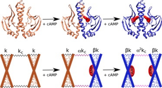
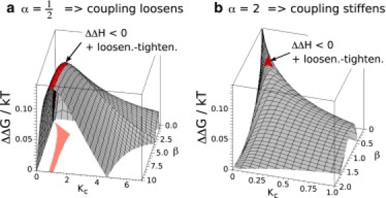
The role of conformational dynamics in allosteric signaling of proteins is increasingly recognized as an important and subtle aspect of this ubiquitous phenomenon. Cooperative binding is commonly observed in proteins with twofold symmetry that bind two identical ligands. We construct a coarse-grained model of an allosteric coupled dimer and show how the signal can be propagated between the distant binding sites via change in slow global vibrational modes alone. We demonstrate that modulation on substrate binding of as few as 5-10 slow modes can give rise to cooperativity observed in biological systems and that the type of cooperativity is given by change of interaction between the two monomers upon ligand binding. To illustrate the application of the model, we apply it to a challenging test case: the catabolite activator protein (CAP). CAP displays negative cooperativity upon association with two identical ligands. The conformation of CAP is not affected by the binding, but its vibrational spectrum undergoes a strong modification. Intriguingly, the first binding enhances thermal fluctuations, yet the second quenches them. We show that this counterintuitive behavior is, in fact, necessary for an optimal anticooperative system, and captured within a well-defined region of the model's parameter space. From analyzing the experimental results, we conclude that fast local modes take an active part in the allostery of CAP, coupled to the more-global slow modes. By including them into the model, we elucidate the role of the modes on different timescales. We conclude that such dynamic control of allostery in homodimers may be a general phenomenon and that our model framework can be used for extended interpretation of thermodynamic parameters in other systems.


Figures

Figure 1
Figure 1
Residues 1–138 of crystallographic structure of CAP (PDB entry 1G6N) binding the ligand cAMP (red) and a sketch of the corresponding coarse-grained model of the system. The large X represents the backbone of one subunit whose one internal mode is simulated by a scissorlike movement of the rods. The little protrusions represent small structures moving fast relative to the slow scissorlike motion of the rods. The internal mode of each subunit and the coupling is defined by the elastic constant k and kc, respectively. The constants are altered upon binding by factors α and β.
Figure 2
Figure 2
(a and b) Allosteric free energy landscapes for a single slow mode. (c and d) Allosteric free energy landscape for one (blue), two (yellow) and three (red) slow modes. The plane ΔΔG = 0 is shown to highlight the border between positive (ΔΔG < 0) and negative cooperativity (ΔΔG > 0).
Figure 3
Figure 3
Four regions with different change in fluctuations mapped onto the ΔΔG landscape for α = 1/2 (a) and α = 2 (b). Color code: In the red region, the loosening-tightening effect is observed. The fluctuations of the doubly liganded system are smaller than that of the apo-protein. The blue region is characterized by the weak loosening-tightening effect, whereby the doubly liganded system moves more than the apo-protein, but less than the 1:1 system. The green region is characterized by sequential stiffening of the protein upon binding. In the gray region, each binding increases the fluctuations. The green region for α > 1 is hidden behind the peak in this view.
Figure 4
Figure 4
Cross-correlation map, Λij, between residue i and j, for three ligation states of CAPN, obtained from the Gaussian network model implemented on the webserver iGNM. A pair subjected to a fully correlated motion (Λij = 1) is colored dark red, fully anticorrelated motions (Λij) are not present, and moderately correlated motions are colored dark blue. cAMP binding disturbs correlations in the liganded monomer (top-left corner of the middle picture) but introduces correlation between the central helices and the liganded monomer. Binding of the second cAMP reestablishes symmetry in the motion pattern and removes correlations of the central helices to the β-sheet structures. Main parts of the secondary structure of CAP are shown above the APO-CAP map; α-helices are represented as magenta cylinders, and β-sheets as gray rectangles.
Figure 5
Figure 5
The allosteric free energy landscapes in the case in which a single slow mode is coupled to a set of identical fast modes, for α = 1/2, 2 with the area displaying the loosening-tightening effect plus ΔΔH < 0 highlighted in red. The projection of the area into the Kc-β plane is shown in orange.

Similar articles

Cited by

References

    1. Kern D., Zuiderweg E.R. The role of dynamics in allosteric regulation. Curr. Opin. Struct. Biol. 2003;13:748–757. - PubMed
    1. Popovych N., Sun S., Kalodimos C.G. Dynamically driven protein allostery. Nat. Struct. Mol. Biol. 2006;13:831–838. - PMC - PubMed
    1. Yonetani T., Laberge M. Protein dynamics explain the allosteric behaviors of hemoglobin. Biochim. Biophys. Acta. 2008;1784:1146–1158. - PMC - PubMed
    1. Tsai C.J., del Sol A., Nussinov R. Allostery: absence of a change in shape does not imply that allostery is not at play. J. Mol. Biol. 2008;378:1–11. - PMC - PubMed
    1. Goodey N.M., Benkovic S.J. Allosteric regulation and catalysis emerge via a common route. Nat. Chem. Biol. 2008;4:474–482. - PubMed

Publication types

LinkOut - more resources