. 2010 May 19;98(10):2327-36.
doi: 10.1016/j.bpj.2010.01.046.

ADF/cofilin binds phosphoinositides in a multivalent manner to act as a PIP(2)-density sensor

Affiliations

ADF/cofilin binds phosphoinositides in a multivalent manner to act as a PIP(2)-density sensor

Hongxia Zhao et al. Biophys J. .

Abstract

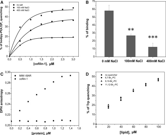
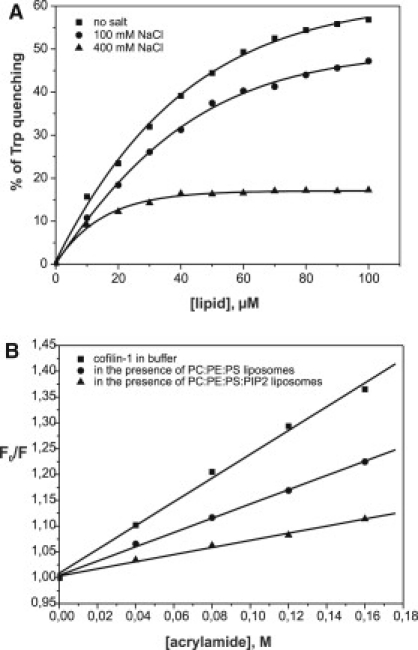
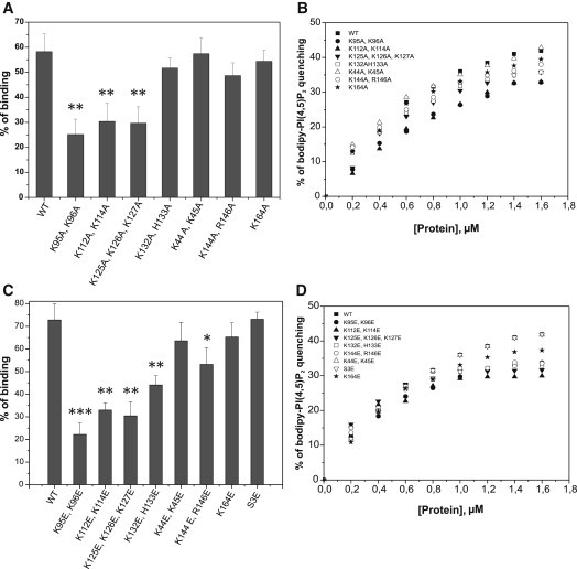
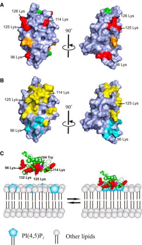
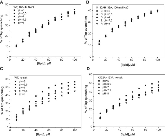
Actin-depolymerizing-factor (ADF)/cofilins have emerged as key regulators of cytoskeletal dynamics in cell motility, morphogenesis, endocytosis, and cytokinesis. The activities of ADF/cofilins are regulated by membrane phospholipid PI(4,5)P(2) in vitro and in cells, but the mechanism of the ADF/cofilin-PI(4,5)P(2) interaction has remained controversial. Recent studies suggested that ADF/cofilins interact with PI(4,5)P(2) through a specific binding pocket, and that this interaction is dependent on pH. Here, we combined systematic mutagenesis with biochemical and spectroscopic methods to elucidate the phosphoinositide-binding mechanism of ADF/cofilins. Our analysis revealed that cofilin does not harbor a specific PI(4,5)P(2)-binding pocket, but instead interacts with PI(4,5)P(2) through a large, positively charged surface of the molecule. Cofilin interacts simultaneously with multiple PI(4,5)P(2) headgroups in a cooperative manner. Consequently, interactions of cofilin with membranes and actin exhibit sharp sensitivity to PI(4,5)P(2) density. Finally, we show that cofilin binding to PI(4,5)P(2) is not sensitive to changes in the pH at physiological salt concentration, although the PI(4,5)P(2)-clustering activity of cofilin is moderately inhibited at elevated pH. Collectively, our data demonstrate that ADF/cofilins bind PI(4,5)P(2) headgroups through a multivalent, cooperative mechanism, and suggest that the actin filament disassembly activity of ADF/cofilin can be accurately regulated by small changes in the PI(4,5)P(2) density at cellular membranes.


Figures

Figure 1
Figure 1
Cofilin-1 is a PI(4,5)P2-density sensor. (A) A vesicle cosedimentation assay shows that the binding of cofilin-1 to membranes is dependent on the PI(4,5)P2 density. The sigmoidal binding isotherm demonstrates that cofilin-1 binds PI(4,5)P2 cooperatively when the density of PI(4,5)P2 in the membrane is augmented. POPE and POPS were included at 20%. PIP2 was included at 0%, 2%, 4%, 6%, 8%, 10%, and 20%, and the POPC concentration was varied accordingly. For 0% PIP2, vesicles composed of POPC/POPE/POPS (6:2:2) were used. The final concentrations of cofilin-1 and liposomes were 2 μM and 500 μM, respectively, in 20 mM Hepes, pH 7.5 buffer. Each data point represents the mean of triplicate measurements, with the error bars indicating ±SD. (B) Membrane binding of cofilin-1 was also examined by quenching of Trp fluorescence. The percentage of quenching shows a sigmoidal PI(4,5)P2-binding isotherm, revealing that cofilin-1 binds PI(4,5)P2 cooperatively and that the interaction is dependent on PI(4,5)P2 density. The cofilin-1 concentration was 1 μM in 20 mM Hepes, 100 mM NaCl, pH 7.5, and total lipid concentration was 140 μM. Lipid composition was as described in panel A. (C) PI(4,5)P2 inhibits the binding of cofilin-1 to actin filaments, and inhibition is PI(4,5)P2-density dependent. Pyrene-labeled actin (20 μM) was polymerized for 30 min in F-buffer (2 mM MgCl2, 1 mM ATP, and 100 mM KCl). The actin filaments were diluted to 1 μM in 20 mM Hepes, 100 mM NaCl, pH 7.5, for fluorescence measurements. Liposomes with different PI(4,5)P2 densities (buffer alone was used as control) were incubated with cofilin-1 for 10 min before addition of the actin filament solution. Pyrene fluorescence was quenched upon cofilin-1 binding to actin filaments. PI(4,5)P2 containing lipsomes inhibited the binding of cofilin-1 to actin filaments, as detected by the decreased quenching of pyrene fluorescence. The final cofilin-1 concentration in the assay was 0.2 μM.
Figure 2
Figure 2
Cofilin simultaneously binds multiple PI(4,5)P2 molecules on the membrane surface and does not insert into the lipid bilayer. (A) Quenching of bodipy-TMR-PI(4,5)P2 demonstrates that cofilin-1 sequesters PI(4,5)P2 on the membrane, and this activity is salt-sensitive. The lipid composition in this assay was POPC/POPE/POPS/PIP2/bodipy-TMR-PI(4,5)P2 (50:20:20:9.5:0.5) and the total lipid concentration was 40 μM. Salt inhibits PI(4,5)P2 clustering by cofilin-1, suggesting electrostatic interactions between cofilin-1 and PI(4,5)P2. (B) A vesicle cosedimentation assay reveals that the cofilin-1-PI(4,5)P2 interaction is inhibited in the presence of high salt, indicative of electrostatic interactions between cofilin-1 and PI(4,5)P2. The assay was carried out under the same conditions (excluding the variation in the salt concentration) as in Fig. 1A. Each data point represents the mean of three independent assays, with the error bars indicating ±SD. A t-test was used to analyze p-values (∗∗∗p < 0.001, ∗∗p < 0.01). (C) Cofilin-1 does not display detectable effects on DPH anisotropy, suggesting that cofilin-1 does not insert into the lipid bilayer upon membrane binding. The I-BAR domain of missing-in-metastasis protein (36) was used as a positive control in this assay. The lipid composition in the assay was POPC/POPE/POPS/PIP2 (5:2:2:1, DPH is incorporated into liposomes at a molar ratio of 1:500) and the total lipid concentration was 40 μM. The assay was carried out in 20 mM Hepes, 100 mM NaCl, pH 7.5. (D) Trp fluorescence of cofilin-1 is not affected by lipids that are brominated at different positions along the acyl chains, indicating that cofilin-1 does not interact with the acyl chains of the lipids. The lipid composition in this assay was POPC/POPE/POPS/PIP2/brominated phosphatidylcholine (2:2:2:1:3). The cofilin-1 concentration was 1 μM.
Figure 3
Figure 3
Trp fluorescence assay shows that cofilin-1 binds PI(4,5)P2, and salt inhibits the interaction. (A) PI(4,5)P2 binding quenches the Trp fluorescence of cofilin-1, suggesting that the environment of Trp-104 is altered due to the binding. Salt inhibits the cofilin-1-PI(4,5)P2 interaction, as detected by the decreased quenching of Trp fluorescence. The final concentration of cofilin-1 in this assay was 1 μM and the lipid composition was POPC/POPE/POPS/PIP2 (5:2:2:1). (B) Stern-Volmer plots for the quenching of cofilin-1 Trp fluorescence by acrylamide in buffer and in the presence of liposomes. Quenching of Trp fluorescence by acrylamide was diminished in the presence of liposomes, indicating that Trp-104 is protected by lipid binding. The final concentrations of cofilin-1 and liposomes were 1 μM and 140 μM, respectively, in 20 mM Hepes, 100 mM NaCl, pH 7.5.
Figure 4
Figure 4
Multiple sequence alignment of selected ADF/cofilins. The residues mutated in mouse cofilin-1 in this study are indicated by lines above the sequences. The mutated residues that displayed severe (red) or moderate (yellow) defects in the PI(4,5)P2-binding assays are highlighted. The conserved Trp, Trp-104, is also highlighted (black).
Figure 5
Figure 5
Determination of the PI(4,5)P2-binding site of cofilin-1. (A) Vesicle cosedimentation assay for examining the effects of various cofilin-1 mutants (alanine substitution) on its PI(4,5)P2-binding activity. This assay suggests that the positively charged clusters K95, K96; K112, K114; and K125, K126, K127 play important roles in PI(4,5)P2 binding by cofilin-1. The assay was carried out under the same conditions described in Fig. 1A. Each data point represents the mean value of three measurements, with the error bars indicating ±SD. A t-test was applied to analyze p-values (∗∗p < 0.01). (B) A bodipy-TMR-PI(4,5)P2 quenching experiment to examine the effects of various cofilin-1 mutants (alanine substitution) for PI(4,5)P2 clustering. This assay revealed the importance of the three positively charged clusters (K95, K96; K112, K114; and K125, K126, K127) in PI(4,5)P2 interactions with cofilin-1. The lipid composition in the assay was POPC/POPE/POPS/PIP2 (5:2:2:1) and the total lipid concentration was 40 μM. Each data point represents the mean of triplicate measurements. (C) The vesicle cosedimentation assay with cofilin-1 mutants (glutamate substitution) suggests that K95, K96; K112, K114; and K125, K126, K127 are the most critical residues in PI(4,5)P2 binding. Each data point represents the mean of triplicate measurements, with the error bars indicating ±SD. A t-test was applied to analyze p-values (∗∗∗p < 0.001, ∗∗p < 0,01, p < 0,05). (D) In similarity to the vesicle cosedimentation assay, the bodipy-TMR-PI(4,5)P2 quenching experiment with cofilin-1 mutants (glutamate substitution) also revealed the importance of the same positively charged clusters (K95, K96; K112, K114; and K125, K126K, 127) on PI(4,5)P2 binding. Each data point represents the mean of triplicate measurements.
Figure 6
Figure 6
A schematic model for the interaction of cofilin with PI(4,5)P2-containing membranes. (A) Space-filling model of the molecular surface of human cofilin-1. The most critical residues in PI(4,5)P2 binding are colored in red and other positively charged residues contributing to PI(4,5)P2 binding are in orange. The mutated residues that did not display effects on PI(4,5)P2 binding are in green. The view on the right represents a 90° clockwise rotation around the long axis of the protein. (B) G-actin and F-actin binding sites of cofilin. Residues that are critical for ADF/cofilin interactions with G-actin and F-actin are in yellow (21,34,41). Residues that are critical for interaction with F-actin, but not G-actin, are in cyan (42–44). The view on the right represents a 90° clockwise rotation around the long axis of the protein. (C) A schematic model for the interaction of cofilin-1 with PI(4,5)P2-rich membranes. The residues that are critical for PI(4,5)P2 binding are indicated in red and PI(4,5)P2 headgroups are indicated in blue. Trp-104 is indicated in magenta. Cofilin-1 simultaneously binds multiple PI(4,5)P2 headgroups through its large, positively charged interface, and thus induces clustering of PI(4,5)P2 on the membrane.
Figure 7
Figure 7
The pH dependency of cofilin-1 binding to PI(4,5)P2. (A and B) PI(4,5)P2 binding quenches the Trp fluorescence of cofilin-1. Based on this assay, the binding of wild-type cofilin-1 (A) and mutant K132A, H133A (B) to PI(4,5)P2-rich membranes is not pH-sensitive under physiological ionic conditions. The final concentration of cofilin-1 in this assay was 1 μM in a total volume of 100 μL of 20 mM Hepes, 100 mM NaCl, pH 7.5. (C and D) PI(4,5)P2 binding of wild-type cofilin-1 (C) is sensitive to changes in pH in the absence of NaCl. However, the binding of mutant K132A, H133A to PI(4,5)P2-rich membranes was less sensitive to changes in pH in the absence of NaCl (D). The final concentration of cofilin-1 was 1 μM in a total volume of 100 μL of 20 mM Hepes, pH 7.5. In both assays the lipid composition was POPC/POPE/POPS/PIP2 (5:2:2:1).
Figure 8
Figure 8
The pH dependency of PI(4,5)P2 clustering by cofilin-1. (A and B) The bodipy-TMR-PI(4,5)P2 quenching assay reveals that PI(4,5)P2 clustering by wild-type cofilin-1 (A) displays moderate pH sensitivity, whereas the K132A, H133A mutant (B) shows no detectable pH sensitivity at physiological ion strength. The final concentration of liposomes was 40 μM in a total reaction volume of 100 μL in 20 mM Hepes, 100 mM NaCl, pH 7.5. The lipid composition was POPC/POPE/POPS/PIP2/Bodipy-TMR-PIP2 (50:20:20:9.5:0.5). The K132A, H133A mutant showed no pH dependency in its ability to cluster PI(4,5)P2, suggesting that His-133 indeed contributes to the pH sensitivity of the PI(4,5)P2-clustering activity of cofilin-1.

Similar articles

Cited by

References

    1. Bamburg J.R., Wiggan O.P. ADF/cofilin and actin dynamics in disease. Trends Cell Biol. 2002;12:598–605. - PubMed
    1. Friedl P., Gilmour D. Collective cell migration in morphogenesis, regeneration and cancer. Nat. Rev. Mol. Cell Biol. 2009;10:445–457. - PubMed
    1. Pollard T.D. The cytoskeleton, cellular motility and the reductionist agenda. Nature. 2003;422:741–745. - PubMed
    1. Paavilainen V.O., Bertling E., Lappalainen P. Regulation of cytoskeletal dynamics by actin-monomer-binding proteins. Trends Cell Biol. 2004;14:386–394. - PubMed
    1. Zebda N., Bernard O., Condeelis J.S. Phosphorylation of ADF/cofilin abolishes EGF-induced actin nucleation at the leading edge and subsequent lamellipod extension. J. Cell Biol. 2000;151:1119–1128. - PMC - PubMed

Publication types

MeSH terms