. 2010 Aug 17;146(1):144-51.
doi: 10.1016/j.jconrel.2010.05.003. Epub 2010 May 18.

Endothelial delivery of antioxidant enzymes loaded into non-polymeric magnetic nanoparticles

Affiliations

Endothelial delivery of antioxidant enzymes loaded into non-polymeric magnetic nanoparticles

Michael Chorny et al. J Control Release. .

Abstract

Antioxidant enzymes have shown promise as a therapy for pathological conditions involving increased production of reactive oxygen species (ROS). However the efficiency of their use for combating oxidative stress is dependent on the ability to achieve therapeutically adequate levels of active enzymes at the site of ROS-mediated injury. Thus, the implementation of antioxidant enzyme therapy requires a strategy enabling both guided delivery to the target site and effective protection of the protein in its active form. To address these requirements we developed magnetically responsive nanoparticles (MNP) formed by precipitation of calcium oleate in the presence of magnetite-based ferrofluid (controlled aggregation/precipitation) as a carrier for magnetically guided delivery of therapeutic proteins. We hypothesized that antioxidant enzymes, catalase and superoxide dismutase (SOD), can be protected from proteolytic inactivation by encapsulation in MNP. We also hypothesized that catalase-loaded MNP applied with a high-gradient magnetic field can rescue endothelial cells from hydrogen peroxide toxicity in culture. To test these hypotheses, a family of enzyme-loaded MNP formulations were prepared and characterized with respect to their magnetic properties, enzyme entrapment yields and protection capacity. SOD- and catalase-loaded MNP were formed with average sizes ranging from 300 to 400 nm, and a protein loading efficiency of 20-33%. MNP were strongly magnetically responsive (magnetic moment at saturation of 14.3 emu/g) in the absence of magnetic remanence, and exhibited a protracted release of their cargo protein in plasma. Catalase stably associated with MNP was protected from proteolysis and retained 20% of its initial enzymatic activity after 24h of exposure to pronase. Under magnetic guidance catalase-loaded MNP were rapidly taken up by cultured endothelial cells providing increased resistance to oxidative stress (62+/-12% cells rescued from hydrogen peroxide induced cell death vs. 10+/-4% under non-magnetic conditions). We conclude that non-polymeric MNP formed using the controlled aggregation/precipitation strategy are a promising carrier for targeted antioxidant enzyme therapy, and in combination with magnetic guidance can be applied to protect endothelial cells from oxidative stress mediated damage. This protective effect of magnetically targeted MNP impregnated with antioxidant enzymes can be highly relevant for the treatment of cardiovascular disease and should be further investigated in animal models.


Figures

Figure 1
Figure 1
Physicochemical properties of magnetic nanoparticles. A. TEM image of MNP. B. MNP size distribution measured from multiple TEM images analyzed with ImageJ software. C. MNP size distribution measured by dynamic light scattering. D. Hysteresis curve of MNP determined using alternating gradient magnetometer. The closed loop through the origin indicates perfect superparamagnetic behavior.
Figure 2
Figure 2
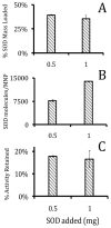
MNP loaded with SOD. A. % SOD activity retained relative to mass added. B. % Mass of SOD loaded relative to mass SOD added. C. Calculated number of SOD molecules per particle based on SOD mass loading.
Figure 3
Figure 3
Catalase loading and catalytic activity as a function of the enzyme mass addition. A. Catalase loading versus input measured by radiotracing of 125I-catalase shows a near-linear relationship. B. Activity of loaded catalase as measured by degradation of hydrogen peroxide absorbance at 242 nm over time reaches saturation at ~1.25 mg.
Figure 4
Figure 4
Release of catalase from MNP. Catalase-loaded MNP were incubated in plasma or aqueous glucose solution (5% w/v) at 37°C, and the amount of 125I-catalase in the release medium was measured over time by radiotracing after MNP centrifugation. –●– Catalase release in aqueous glucose solution. –○– Catalase release in whole heparinized mouse plasma. Note the biphasic release pattern in plasma with a rapid initial release phase followed by a plateau.
Figure 5
Figure 5
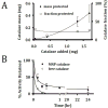
Protection of catalase mass and activity from proteolysis. A. Mass of MNP-encapsulated catalase protected from proteolysis upon exposure to 0.2 wt% Pronase at 37°C for 1 hr vs. catalase input. B. Protection of the enzymatic activity of catalase loaded into MNP vs. free catalase upon exposure to 0.2 wt% Pronase at 37°C.
Figure 6
Figure 6
Magnetic delivery of MNP to cultured bovine aortic endothelial cells. MNP formed with Dylight 488-labeled catalase were applied to cells with or without an exposure to a high gradient magnetic field (10 min), and the cell uptake of MNP and catalase were observed microscopically 4 hr post treatment using the bright field and FITC fluorescent channels, respectively, in comparison to untreated cells (A–F). Original magnification ×100. Note the large number of perinuclearly localized MNP in cells treated under magnetic onditions, which is paralleled by a similar cellular distribution pattern of the fluorescent labeled enzyme. Also note the absence of visible MNP or enzyme in non-magnetic control cells. Quantitative assay of cell uptake of blank and catalase-loaded MNP revealed similar patterns with significantly larger amounts of MNP internalized by 4 hr after magnetic delivery (G). Cell viability measured 24 hr post treatment with MNP under magnetic conditions was not adversely affected (H).
Figure 7
Figure 7
Protection of HUVEC from oxidative stress through magnetic delivery of catalase loaded MNP. Viability of cells pretreated with MNP and exposed to 10 mM hydrogen peroxide for 5 hour was determined fluorimetrically after staining with Calcein AM. A. Untreated cells used as a reference. B. Cells exposed to hydrogen peroxide only (no protection). C. Cells treated under magnetic conditions with MNP-encapsulated catalase. Original magnification ×100. D. Quantification of the viability of hydrogen peroxide-challenged cells treated with catalase-loaded MNP in the presence of a high gradient magnetic field in comparison to controls.

References

    1. Laurent S, Forge D, Port M, Roch A, Robic C, Vander Elst L, Muller RN. Magnetic iron oxide nanoparticles: synthesis, stabilization, vectorization, physicochemical characterizations, and biological applications. Chem Rev. 2008;108(6):2064–2110. - PubMed
    1. Chorny M, Polyak B, Alferiev IS, Walsh K, Friedman G, Levy RJ. Magnetically driven plasmid DNA delivery with biodegradable polymeric nanoparticles. FASEB J. 2007;21(10):2510–2519. - PMC - PubMed
    1. Namdeo M, Saxena S, Tankhiwale R, Bajpai M, Mohan YM, Bajpai SK. Magnetic nanoparticles for drug delivery applications. J Nanosci Nanotechnol. 2008;8(7):3247–3271. - PubMed
    1. Veiseh O, Gunn JW, Kievit FM, Sun C, Fang C, Lee JS, Zhang M. Inhibition of tumor-cell invasion with chlorotoxin-bound superparamagnetic nanoparticles. Small. 2009;5(2):256–264. - PMC - PubMed
    1. Ma HL, Qi XR, Ding WX, Maitani Y, Nagai T. Magnetic targeting after femoral artery administration and biocompatibility assessment of superparamagnetic iron oxide nanoparticles. J Biomed Mater Res A. 2008;84(3):598–606. - PubMed

Publication types

MeSH terms