. 2010 Jun 15;184(12):6822-32.
doi: 10.4049/jimmunol.0902058. Epub 2010 May 14.

Invariant NKT cell development requires a full complement of functional CD3 zeta immunoreceptor tyrosine-based activation motifs

Affiliations

Invariant NKT cell development requires a full complement of functional CD3 zeta immunoreceptor tyrosine-based activation motifs

Amy M Becker et al. J Immunol. .

Abstract

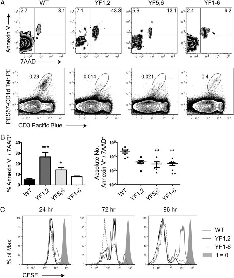
Invariant NKT (iNKT) cells regulate early immune responses to infections, in part because of their rapid release of IFN-gamma and IL-4. iNKT cells are proposed to reduce the severity of Lyme disease following Borrelia burgdorferi infection. Unlike conventional T cells, iNKT cells express an invariant alphabeta TCR that recognizes lipids bound to the MHC class I-like molecule, CD1d. Furthermore, these cells are positively selected following TCR interactions with glycolipid/CD1d complexes expressed on CD4+CD8+ thymocytes. Whereas conventional T cell development can proceed with as few as 4/10 CD3 immunoreceptor tyrosine-based activation motifs (ITAMs), little is known about the ITAM requirements for iNKT cell selection and expansion. We analyzed iNKT cell development in CD3 zeta transgenic lines with various tyrosine-to-phenylalanine substitutions (YF) that eliminated the functions of the first (YF1,2), third (YF5,6), or all three (YF1-6) CD3 zeta ITAMs. iNKT cell numbers were significantly reduced in the thymus, spleen, and liver of all YF mice compared with wild type mice. The reduced numbers of iNKT cells resulted from significant reductions in the expression of the early growth response 2 and promyelocytic leukemia zinc finger transcription factors. In the mice with few to no iNKT cells, there was no difference in the severity of Lyme arthritis compared with wild type controls, following infections with the spirochete B. burgdorferi. These findings indicate that a full complement of functional CD3 zeta ITAMs is required for effective iNKT cell development.


Figures

FIGURE 1
FIGURE 1
iNKT cell development requires a full complement of functional CD3 ζ ITAMs. A, The percentage of iNKT cells in the thymus, spleens, and livers of the indicated mice were identified by staining cells with anti-CD3 ε-Pacific Blue in combination with PBS57-loaded CD1d tetramer-PE and then analyzing the cells by flow cytometry. iNKT cells are represented in oval gates. The percentage (B) and absolute number (C) of iNKT cells from WT, YF1,2, YF5,6, and YF1–6 mice were compared using at least five mice per group. D, The absolute number of thymocytes and splenocytes from at least five mice per group are shown. Data are consistent from four independent experiments. **p < 0.001; ***p = 0.0001; values were generated by Kruskal–Wallis test.
FIGURE 2
FIGURE 2
iNKT cell development is blocked at an early stage of thymopoiesis in mice lacking one or more functional CD3 ζ ITAMs. Thymocytes or splenocytes from wild type, YF1,2, YF5,6, and YF1–6 mice were stained with anti-CD3 ε-Pacific Blue, PBS57-CD1d tetramer-PE, anti-HSA FITC, -CD44 PeCy7, –NK1.1 allophycocyanin, and –CD4 PerCp-Cy5,5. A, The percentage of HSAlo iNKT cells was analyzed by gating on HSAlo PBS57-CD1d tetramer+ lymphocytes (left gate). The forward scatter and side scatter profiles of the cells analyzed in the upper panel are shown in the lower panel. B, Stage I, II, and III iNKT cells were analyzed in the thymus and spleen of indicated mice, as indicated in corresponding quadrants labeled I, II, and III. Quadrants represent CD3intermediate CD1d tetramer+ HSAlo cells. C, The percentage of CD4 SP and DN iNKT cells was analyzed on CD3intermediate CD1d tetramer+ HSAlo cells. Graph represents the ratio of CD4 to DN iNKT cells. Three to seven mice were analyzed per group. Data are representative of three independent experiments. Statistics were generated by Kruskal–Wallis test. ***p < 0.0001; **p < 0.001.
FIGURE 3
FIGURE 3
Mice lacking two or more CD3 ζ ITAMs have an increase in cell death compared with controls. A, Thymic iNKT cells from the indicated mice were stained with anti-CD3 ε Pacific Blue, PBS57-loaded CD1d tetramer PE, annexin V allophycocyanin, and 7AAD and then analyzed by flow cytometry. Annexin V and 7AAD staining was analyzed on CD3intermediate CD1d tetramer+ iNKT cells. Numbers represent the percentage of cells in each quadrant. B, Graph indicates the percentage or absolute numbers of iNKT cells that are annexin V+ 7AAD+ using at least six mice per group pooled from three independent experiments. Statistics were generated by Kruskal–Wallis test. ***p < 0.0001; **p < 0.001. C, Proliferative responses of iNKT cells. Cells from the various mice were labeled with CSFE and stimulated with α-galactosylceramide for the indicated times. The cells were subsequently washed and analyzed by flow cytometry. Data are representative of three independent experiments.
FIGURE 4
FIGURE 4
iNKT cells from mice lacking one or more functional CD3 ζ ITAMs have reduced expression of multiple transcription factors. A, Total RNA was isolated from thymocytes obtained from wild type, YF1,2, YF5,6, YF1–6, and Jα18 knockout mice (Jα18KO). Equivalent amounts of RNA were used for real-time RT-PCR reactions with primers specific for Vα14Jα18 rearrangements. Primers for Cα were used as controls. Samples were run in triplicate, with GAPDH included as an internal control. Vα14Jα18 and Cα expression was first normalized to GAPDH. Data represent relative expression compared with wild type controls. B, iNKT cells were isolated from pooled splenocytes using magnetic bead depletion and cell sorting with a high-speed cell sorter. Total RNA was isolated from the purified iNKT cells obtained from wild type, YF1,2, YF5,6, and YF1–6 mice. The RNA was used in real-time RT-PCR reactions with primers specific for Egr1, Egr2, Egr3, c-Myc, and PLZF, with GAPDH included as a control for the RT-PCR reactions. The relative expression of each transcription factor was calculated from five independent real-time experiments using two or three independent cell sorts. The statistical analyses were not included in the YF1,2 and YF5,6 samples, because the amplification for Egr2 and PLZF was below the critical threshold in three and four reactions, respectively. In those samples in which the Ct values were detected, the values were normalized to GAPDH. Data represent the relative expression of the transcription factors in the YF line compared with wild type mice. n is the number of successful PCR reactions from a total of five or six attempts. C, Thymocytes were obtained from the indicated mice and aliquots were stained with fluorescently labeled mAbs against CD4 PercpCy5.5, CD8 PeCy7, CD1d, Ly108, or CD150 PE. Histograms represent D thymocytes. Data are representative of two independent experiments.
FIGURE 5
FIGURE 5
iNKT cells are not necessary to prevent inflammatory arthritis following B. burgdorferi infection. Six-week-old mice were inoculated intradermally with 1.2–1.5 × 10⁶ B. burgdorferi spirochetes. Right and left tibiotarsal joints were isolated and paraffin embedded, and 7-μm sections were cut and stained with H&E. A and B, Digital pictures taken of tibiotarsal joints from indicated mice. C and D, Joints were scored for arthritis on a scale 0–3 indicating no, slight, moderate, or severe inflammation, respectively. Bar graphs indicated the mean arthritis score at 2 or 6 wk postinfection. Three sham and six to nine infected mice were analyzed per group at each time point. All images were acquired with a ×4 objective magnification.
FIGURE 6
FIGURE 6
iNKT cells are not necessary to prevent inflammatory aortitis following B. burgdorferi infection. Five eight-week-old mice were prebled and inoculated intradermally with 1.5 × 10⁶ B. burgdorferi spirochetes. Two weeks later, all the control and infected mice were bled. The hearts were isolated and paraffin embedded, and 7-μm sections were cut and stained with H&E. A, Aortitis was determined using a blinded scoring system in which inflammation was defined on a scale of 0–4 indicating no, slight, mild, moderate, or severe inflammation, respectively. Bar graphs indicated the mean aortitis 2 wk postinfection. Three shams and a minimum of five infected mice were analyzed per group. There was no statistically significant difference in the mice when analyzed by Kruskal– Wallis tests. B, Serum from the indicated uninfected and B. burgdorferi-infected mice was serially diluted as indicated and used in an ELISA assay to test for reactivity to B. burgdorferi Ags.

Similar articles

Cited by

References

    1. Bendelac A, Savage PB, Teyton L. The biology of NKT cells. Annu. Rev. Immunol. 2007;25:297–336. - PubMed
    1. Tupin E, Kinjo Y, Kronenberg M. The unique role of natural killer T cells in the response to microorganisms. Nat. Rev. Microbiol. 2007;5:405–417. - PubMed
    1. Godfrey DI, MacDonald HR, Kronenberg M, Smyth MJ, Van Kaer L. NKT cells: what's in a name? Nat. Rev. Immunol. 2004;4:231–237. - PubMed
    1. Godfrey DI, Stankovic S, Baxter AG. Raising the NKT cell family. Nat. Immunol. 2010;11:197–206. - PubMed
    1. Kumar H, Belperron A, Barthold SW, Bockenstedt LK. Cutting edge: CD1d deficiency impairs murine host defense against the spirochete, Borrelia burgdorferi. J. Immunol. 2000;165:4797–4801. - PubMed

Publication types