. 2010 Jun 15;184(12):7010-21.
doi: 10.4049/jimmunol.0904133. Epub 2010 May 14.

Human neuronal cells possess functional cytoplasmic and TLR-mediated innate immune pathways influenced by phosphatidylinositol-3 kinase signaling

Affiliations

Human neuronal cells possess functional cytoplasmic and TLR-mediated innate immune pathways influenced by phosphatidylinositol-3 kinase signaling

Daniel C Peltier et al. J Immunol. .

Abstract

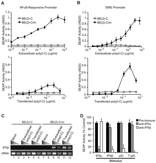
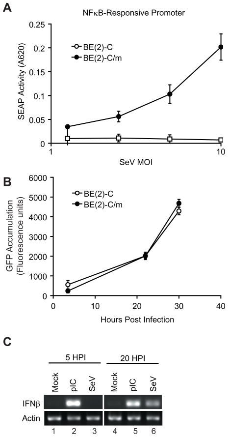
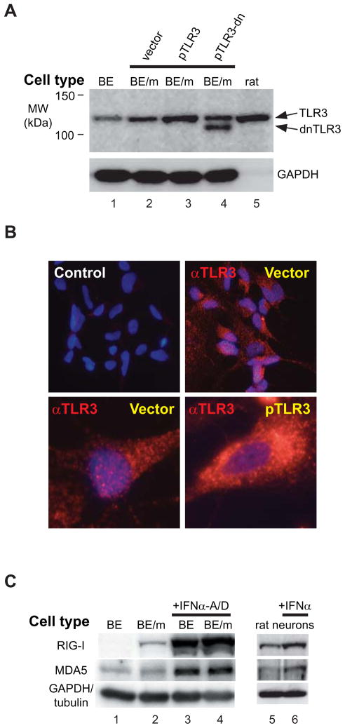
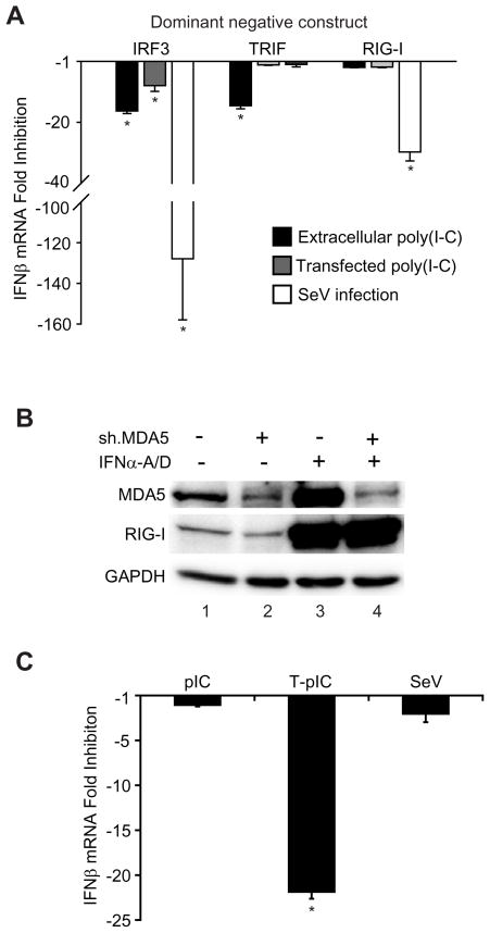
Innate immune pathways are early defense responses important for the immediate control and eventual clearance of many pathogens, where signaling is initiated via pattern recognition receptor (PRR)-mediated events that occur in a ligand- and cell-type specific manner. Within CNS neurons, innate immune pathways are likely crucial to control pathogens that target these essential yet virtually irreplaceable cells. However, relatively little is known about the induction and regulation of neuronal PRR signaling. In this report, we used human neuronal cell lines and primary rat neuronal cultures to examine PRR expression and function. We found that several innate immune receptor ligands, including Sendai virus, the dsRNA mimetic polyinosinic-polycytidylic acid, and LPS all activated differentiation-dependent neuronal innate immune pathways. Functional genetic analyses revealed that IFN regulatory factor 3-mediated pathways that resulted in IFN-beta transcriptional upregulation were activated in cultured human neuronal cells by the PRRs TLR3, MDA5, or RIG-I in a ligand-specific manner. Furthermore, genome-wide transcriptional array and targeted genetic and pharmacologic analyses identified PI3K signaling as crucial for the induction of innate immune pathways in neurons. These results indicate that human neuronal cells possess specific and functional PRR pathways essential for the effective induction of innate immune responses, and suggest that neurons can play an active role in defense against neurotropic pathogens.


Figures

FIGURE 1
FIGURE 1
Poly(I-C) activates NFκB and ISRE promoters and induces IFNβ production in human neuronal cells. Similar numbers of NFκB (A) or ISRE (B) promoter-reporter cells were stimulated with increasing amounts of extracellular poly(I-C) (upper graphs) or transfected poly(I-C) (lower graphs), and SEAP reporter activity in culture supernatants was measured 20 h after stimulation. (C) BE(2)-C (lanes 1–6) or BE(2)-C/m (lanes 7–12) cells were stimulated with 100 μg/ml extracellular poly(I-C) (lanes 3, 4, 9, and 10) or 700 ng/ml transfected poly(I-C) (lanes 5, 6, 11, and 12) for 10 h, and IFNβ and rRNA transcript levels were assessed via RT-PCR. Adjacent lanes for individual samples represent results using 10-fold dilutions of cDNA. (D) BE(2)-C/m ISRE reporter cells were stimulated with either 20 IU/ml human leukocyte IFNα, 20 IU/ml human fibroblast IFNβ, 100 μg/ml extracellular poly(I-C) (pIC), or 600 ng/ml transfected poly(I-C) (T-pIC) in the presence of neutralizing IFNα or IFNβ antisera at concentrations capable of neutralizing 4000 IU/ml of IFNα or IFNβ respectively. Results are expressed as the percent SEAP activity compared to control samples incubated with pre-immune serum.
FIGURE 2
FIGURE 2
SeV infection induces a neuronal PRR response. (A) NFκB reporter cells were infected with GFP-tagged SeV using an increasing multiplicity of infection (MOI), and SEAP reporter activity was assessed at 30 hpi. (B) Cells were infected as in A and SeV replication, assessed by GFP fluorescence, was measured. (C) BE(2)-C/m cells were stimulated with 100 μg/ml extracellular poly(I-C) (pIC, lanes 2 and 5) or infected with SeV at an MOI of 10 (lanes 3 and 6), and IFNβ mRNA or rRNA accumulation was assessed via RT-PCR 5 h (lanes 1–3) or 20 h (lanes 4–6) later.
FIGURE 3
FIGURE 3
Human neuronal cells and differentiated rodent neurons express antiviral PRRs. (A) Lysates from BE(2)-C cells (lane 1), differentiated BE(2)-C/m cells (lanes 2–4), or primary rat neurons (lane 5) were immunoblotted for TLR3 levels. To validate antibody specificity, BE(2)-C/m cells were transfected with either empty vector (lane 2) or plasmids expressing wild-type TLR3 (lane 3) or a dominant-negative (dn) TLR3 (lane 4) that contains a TIR domain deletion. The human TLR3 gene encodes a 904 amino acid protein with a predicted MW of 103 kDa, although it is heavily glycosylated. The TLR3 ΔTIR mutant contains a 162 amino acid deletion that reduces the predicted MW by approximately 18 kDa. The GAPDH-specific monoclonal antibody used for immunoblotting cross-reacted poorly with the rat lysate (lane 5), but total protein staining showed that the rat lysate sample contained approximately 2- to 3-fold more total protein than the other lanes (data not shown). (B) Immunofluorescent staining of TLR3 expression in BE(2)-C/m cells. The primary TLR3-specific antibody was excluded during incubation in control cells (upper left image). Nuclei were stained with DAPI (blue), whereas the punctate PE-staining (red) indicates TLR3 expression. Cells in the upper right and lower left images were transfected with empty vector, whereas cells in the lower right image were transfected with a plasmid expressing wild-type TLR3. We also saw a similar punctuate pattern but increased TLR3 signal intensity in cells transfected with the dnTLR3 expression plasmid (data not shown). (C) Lysates from BE(2)-C cells (lanes 1 and 3), differentiated BE(2)-C/m cells (lanes 2 and 4), or primary rat neurons (lanes 5 and 6), were immunoblotted for RIG-I, MDA5, and GAPDH (human) or tubulin (rat) levels. Lysates from cells treated with 100 IU/ml human IFNα-A/D for 6 h (lanes 3 and 4) or 50 IU/ml rat IFNα for 24 h (lane 6) were used as controls to validate the identity of RIG-I and MDA5 as IFN-stimulated genes.
FIGURE 4
FIGURE 4
Human neuronal cells possess functional PRR-mediated innate immune pathways. (A) BE(2)-C/m cells stably overexpressing dominant negative forms of IRF3, TRIF, or RIG-I were stimulated with 100 μg/ml extracellular poly(I-C) or 700 ng/ml transfected poly(I-C) for 10 h, or infected with SeV for 30 h, and IFNβ mRNA levels were measured by quantitative RT-PCR using rRNA transcript levels as the loading control. Results are expressed as the fold-change compared to similarly stimulated cells stably transfected with an empty vector. (B) Lysates from BE(2)-C/m cells stably transfected with plasmids expressing shRNAs targeted against either a control protein (lanes 1 and 3) or MDA5 (lanes 2 and 4) were immunoblotted for MDA5, RIG-I, and GAPDH expression levels. The level of MDA5 suppression in cells expressing an MDA5-specific shRNA was 44.5 ± 7.4% compared to control cells. Live-cell imaging of differentiated cells also demonstrated that greater than 95% of cells expressed the control GFP reporter gene encoded on the shRNA expression plasmid (data not shown). Lysates from cells treated with 1000 IU/ml IFNα-A/D for 12 h (lanes 3 and 4) served as positive controls to validate the specificity of shRNA-mediated knockdown of MDA5 under enhanced expression levels. (C) IFNβ mRNA levels in BE(2)-C/m cells stably expressing an MDA5-targeted shRNA after stimulation with extracellular poly(I-C) (pIC), transfected poly(I-C) (T-pIC), or infected with SeV as described above. Transcript levels were determined by quantitative RT-PCR and results are expressed as the fold-change compared to similarly stimulated cells stably transfected with an shRNA-encoding vector targeting an irrelevant control protein. *p-value < 0.05.
FIGURE 5
FIGURE 5
Neuronal response to poly(I-C) is mediated by PI3K. (A) BE(2)-C/m ISRE (upper graph) and NFκB (lower graph) promoter-driven reporter cells were treated with an increasing concentration of LY294002, stimulated with 100 μg/ml extracellular poly(I-C) (pIC), 700 ng/ml transfected poly(I-C) (T-pIC), 100 IU/ml IFNα-A/D, 50 ng/ml TNFα, or 500 ng/ml LPS, and SEAP reporter activity was measured 24 h later. Results are expressed as the percentage of SEAP activity compared to DMSO-treated controls. TNFα and LPS were used as controls only with NFκB promoter-driven reporter cells as the ISRE reporter cells did not respond either stimuli even in the absence of LY294002 (see Table I and data not shown). (B) Primary rat cortical neurons were treated with either DMSO (lanes 1 and 2) or 10 μM LY294002 (lane 3), stimulated with 50 μg/ml of extracellular poly(I-C) for 8 h (lanes 2 and 3), and IFNβ mRNA levels were assessed by RT-PCR.
FIGURE 6
FIGURE 6
PI3K catalytic subunit p110α mediates human neuronal cell responses to poly(I-C). (A) BE(2)-C/m ISRE reporter cells were treated with increasing concentrations of a selective PI3K p110α (p110a Inhibitor 2), p110β (TGX-221), or p110γ (AS-252424) catalytic subunit inhibitor, stimulated with 100 μg/ml extracellular poly(I-C) (pIC), 700 ng/ml transfected poly(I-C) (T-pIC), or 100 IU/ml IFNα-A/D, and SEAP reporter activity was measured 20 h later. Results are presented as the percent reporter gene activity compared to DMSO-treated controls. (B) BE(2)-C/m cells were treated with 10 μM LY294002, 5 μM p110α Inhibitor 2, or 5 μM AS-252424, stimulated with poly(I-C) as described above, and IFNβ mRNA levels were measured 4 h later by quantitative RT-PCR. *p-values < 0.05.
FIGURE 7
FIGURE 7
The PI3K p110α catalytic subunit mediates a TLR3-dependent response in neuronal cells. (A) Lysates from BE(2)-C/m cells stably transduced with lentiviruses expressing an empty vector (lane 1) or shRNAs targeted against either a control protein (CD14, lane 2) or PI3K p110α (lane 3) were immunoblotted for p110α and GAPDH expression levels. The level of suppression in cells expressing a p110α-specific shRNA was 60.4 ± 4.9% compared to control cells. Live-cell imaging of differentiated cells also demonstrated that greater than 95% of cells expressed the control GFP reporter gene encoded on the shRNA expression plasmid (data not shown). (B) IFNβ mRNA levels in BE(2)-C/m cells stably expressing a p110α-targeted shRNA after stimulation with extracellular poly(I-C) (pIC) or transfected poly(I-C) (T-pIC) as described in Fig. 6. Transcript levels were determined by quantitative RT-PCR and results are expressed as the fold-change compared to similarly stimulated cells stably transfected with an shRNA-encoding vector targeting an irrelevant control protein. *p-value < 0.05.

Similar articles

Cited by

References

    1. Kumar H, Kawai T, Akira S. Pathogen recognition in the innate immune response. Biochem J. 2009;420:1–16. - PubMed
    1. Pichlmair A, Reis e Sousa C. Innate recognition of viruses. Immunity. 2007;27:370–383. - PubMed
    1. Schmidt A, Schwerd T, Hamm W, Hellmuth JC, Cui S, Wenzel M, Hoffmann FS, Michallet MC, Besch R, Hopfner KP, Endres S, Rothenfusser S. 5′-triphosphate RNA requires base-paired structures to activate antiviral signaling via RIG-I. Proc Natl Acad Sci U S A. 2009;106:12067–12072. - PMC - PubMed
    1. Schlee M, Roth A, Hornung V, Hagmann CA, Wimmenauer V, Barchet W, Coch C, Janke M, Mihailovic A, Wardle G, Juranek S, Kato H, Kawai T, Poeck H, Fitzgerald KA, Takeuchi O, Akira S, Tuschl T, Latz E, Ludwig J, Hartmann G. Recognition of 5′ triphosphate by RIG-I helicase requires short blunt double-stranded RNA as contained in panhandle of negative-strand virus. Immunity. 2009;31:25–34. - PMC - PubMed
    1. Pichlmair A, Schulz O, Tan CP, Rehwinkel J, Kato H, Takeuchi O, Akira S, Way M, Schiavo G, Reis e Sousa C. Activation of MDA5 requires higher-order RNA structures generated during virus infection. J Virol. 2009;83:10761–10769. - PMC - PubMed

Publication types

Substances