. 2010 Jun 15;184(12):6807-14.
doi: 10.4049/jimmunol.1000453. Epub 2010 May 17.

Differential roles of IL-2-inducible T cell kinase-mediated TCR signals in tissue-specific localization and maintenance of skin intraepithelial T cells

Affiliations

Differential roles of IL-2-inducible T cell kinase-mediated TCR signals in tissue-specific localization and maintenance of skin intraepithelial T cells

Mingcan Xia et al. J Immunol. .

Abstract

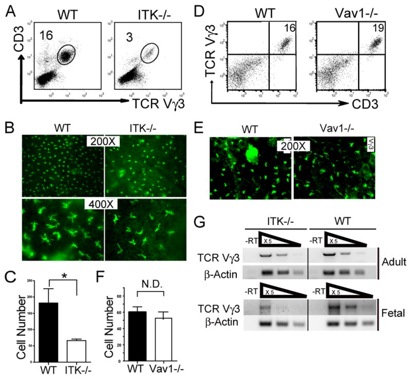
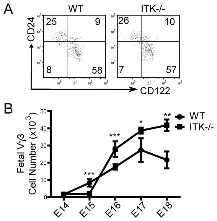
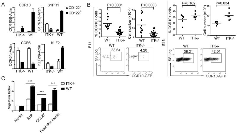
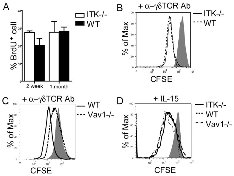
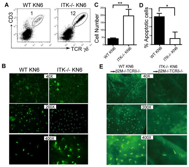
Tissue-specific innate-like gammadelta T cells are important components of the immune system critical for the first line of defense, but mechanisms underlying their tissue-specific development are poorly understood. Our study with prototypical skin-specific intraepithelial gammadeltaT lymphocytes (sIELs) found that among different thymic gammadelta T cell subsets fetal thymic precursors of sIELs specifically acquire a unique skin-homing property after positive selection, suggesting an important role of the TCR selection signaling in "programming" them for tissue-specific development. In this study, we identified IL-2-inducible T cell kinase (ITK) as a critical signal molecule regulating the acquirement of the skin-homing property by the fetal thymic sIEL precursors. In ITK knockout mice, the sIEL precursors could not undergo positive selection-associated upregulation of thymus-exiting and skin-homing molecules sphingosine-1-phosphate receptor 1 and CCR10 and accumulated in the thymus. However, the survival and expansion of sIELs in the skin did not require ITK-transduced TCR signaling, whereas its persistent activation impaired sIEL development by inducing apoptosis. These findings provide insights into molecular mechanisms underlying differential requirements of TCR signaling in peripheral localization and maintenance of the tissue-specific T cells.


Figures

Figure 1
Figure 1
Impaired development of sIELs in ITK−/− but not Vav1−/− mice. A. Skin cell preparations from 6–8 week old ITK−/− and wild type mice were stained with anti-CD3 and Vγ3 antibodies, and analyzed for percentages of the CD3+Vγ3+ population by flow cytometry. One representative of three independent experiments is shown. B and C. Ear epidermal sheets from wild type and ITK−/− mice were stained with fluorescent anti-Vγ3 antibody and observed under a fluorescent microscope (Olympus BX61) for the Vγ3+ sIELs (B), average numbers of which per field at the 200X amplification were plotted (C). Data were obtained from three independent experiments. *P < 0.05. D-F. Flow cytometric and immunofluorescent microscopic analysis of skin Vγ3+ γδ T cells from Vav1−/− mice as performed in the panels A-C except that the Vγ3+ sIELs on the epidermal sheets was visualized under a different fluorescent microscope (Nikon Eclipse TE 300) that has a smaller field at the same 200X amplification (E). Experiments were repeated twice for both flow cytometric and immunofluorescent analyses. Average numbers of sIELs per field obtained in the panel E were plotted in the panel F. N.D: no difference. G. Total RNA from fetal and adult mouse skin was reverse transcribed to cDNA. Serially 5 fold-diluted cDNA were subject to semi-quantitative PCR to determine expression levels of rearranged TCRγ3 gene. β-actin was used as a control. Data shown were obtained from three independent experiments.
Figure 2
Figure 2
Vγ3+ sIEL precursors undergo a normal maturation process but accumulate in the fetal thymus of ITK−/− mice. A. Flow cytometric analysis of CD122 and CD24 expression on gated E16–17 fetal thymic Vγ3+ γδ cells. One representative of three independent experiments is shown. B. Numbers of Vγ3+ γδ T cells in wild type and ITK−/− fetal thymi of different gestation ages. The numbers were calculated based on total numbers of thymocytes and percentages of Vγ3+ cells per thymus. Data presented were means and standard deviations from three to five experiments. * P < 0.05, ** P <0.01, *** P < 0.001.
Figure 3
Figure 3
ITK−/− fetal thymic sIEL precursors exhibit the altered migration molecule expression and defective migration capability. A. Real-time RT-PCR analysis of the expression of indicated molecules in purified CD122+ and CD122 CD3+Vγ3+ cells of E16 wild type and ITK−/− fetal thymi. Data shown were obtained from three independent experiments. B. E14 or16 fetal thymocytes of ITK−/−CCR10+/EGFP and ITK+/−CCR10+/EGFP mice were analyzed for CCR10 (EGFP) expression on Vγ3+ cells. Percentages and numbers of CCR10 (EGFP)+ Vγ3+ cells were shown. Data presented is one representative from at least 6 mice of each genotype. C. In vitro migration of wild type and ITK−/− E16 fetal thymic Vγ3+ γδ T cells to S1P, CCL27 and conditioned medium of fetal skin cultures. The migration index was calculated as a ratio of numbers of Vγ3+ cells migrating into the bottom chamber in presence of attractants vs. medium only. Data shown were obtained from two independent experiments. *** P < 0.001.
Figure 4
Figure 4
ITK−/− sIELs and their fetal thymic precursors have normal proliferation capacities. A. Similar in vivo proliferation rates of wild type and ITK−/− sIELs. Two week or one-month old mice were treated with BrdU for 9 days and then sIELs were isolated and analyzed for BrdU incorporation by flow cytometry. Data presented were means and standard deviations from three to five experiments. B-D. CFSE-labeled E16 fetal thymocytes from ITK−/−, Vav1−/− or wild type mice were stimulated with anti-γδ TCR antibody (1μg/ml, GL4) or IL-15 (50 ng/ml) for 3 days, and analyzed by flow cytometry for the proliferation of CD3+γδ T cells. One representative of three independent experiments was shown.
Figure 5
Figure 5
ITK-mediated TCR/ligand induced signaling in the skin impairs the development of sIELs by promoting their apoptosis. A. Skin cell preparations of KN6 transgenic mice of wild type and ITK−/− backgrounds were analyzed for transgenic Vγ2+ sIELs by flow cytometry. Percentages of the transgenic sIELs were indicated. One representative of three independent experiments is shown. B and C. Ear epidermal sheets of ITK-sufficient and knockout KN6 mice were stained and observed under a fluorescent microscope (Olympus BX61) for transgenic Vγ2+ sIELs (B), average numbers of which per field at the 200x amplification were plotted (C). Data were obtained from three experiments. ** P < 0.01. D. Lower percentages of apoptotic KN6 transgenic sIELs on ITK−/− than wild type background. The percentages of apoptotic sIELs were calculated based on ratios of numbers of apoptotic vs. total sIELs from in situ TUNEL analyses of ear epidermal sheets, as shown in the Supplementary Fig. 3. Data shown were obtained from at least four mice of each genotype in two independent experiments. * P < 0.05. E. The development of KN6 transgenic sIELs in β2m−/−TCRδ −/− recipients from adoptively transferred fetal thymic ITK-sufficient or knockout fetal thymic KN6 transgenic γδ T cells. Ear epidermal sheets of the recipients were analyzed for donor-derived sIELs by in situ immunofluorescent staining (Olympus BX61). Data presented is one representative from three mice of each genotype.

References

    1. Hayday AC. [gamma][delta] cells: a right time and a right place for a conserved third way of protection. Annual review of immunology. 2000;18:975–1026. - PubMed
    1. Raulet DH, Spencer DM, Hsiang YH, Goldman JP, Bix M, Liao NS, Zijstra M, Jaenisch R, Correa I. Control of gamma delta T-cell development. Immunological reviews. 1991;120:185–204. - PubMed
    1. Elbe A, Tschachler E, Steiner G, Binder A, Wolff K, Stingl G. Maturational steps of bone marrow-derived dendritic murine epidermal cells. Phenotypic and functional studies on Langerhans cells and Thy-1+ dendritic epidermal cells in the perinatal period. J Immunol. 1989;143:2431–2438. - PubMed
    1. Havran WL, Allison JP. Origin of Thy-1+ dendritic epidermal cells of adult mice from fetal thymic precursors. Nature. 1990;344:68–70. - PubMed
    1. Mallick-Wood CA, Lewis JM, Richie LI, Owen MJ, Tigelaar RE, Hayday AC. Conservation of T cell receptor conformation in epidermal gammadelta cells with disrupted primary Vgamma gene usage. Science (New York, NY) 1998;279:1729–1733. - PubMed

Publication types

MeSH terms

Substances