. 2010 Jun 15;184(12):7207-18.
doi: 10.4049/jimmunol.0903487. Epub 2010 May 7.

The prostaglandin E2 E-prostanoid 4 receptor exerts anti-inflammatory effects in brain innate immunity

Affiliations

The prostaglandin E2 E-prostanoid 4 receptor exerts anti-inflammatory effects in brain innate immunity

Ju Shi et al. J Immunol. .

Abstract

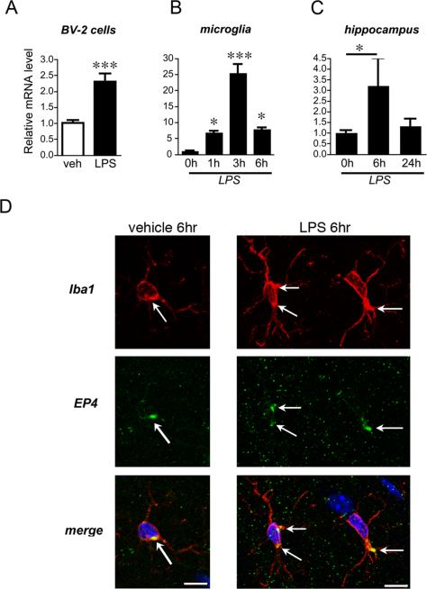
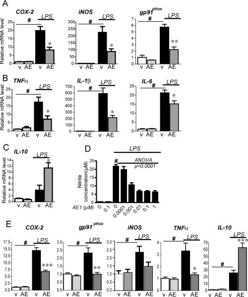
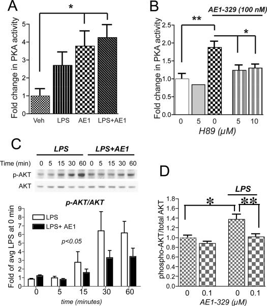
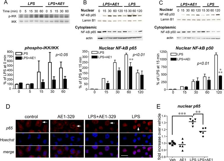
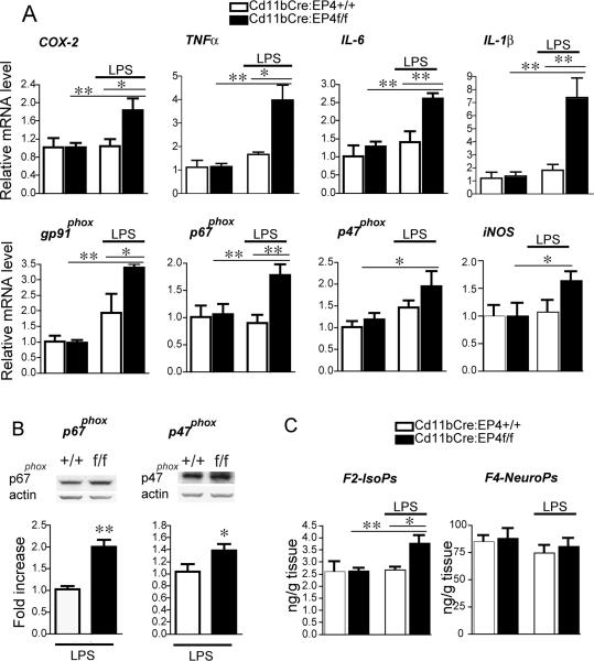
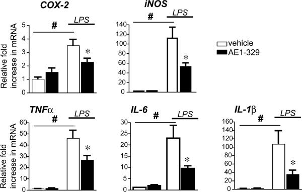
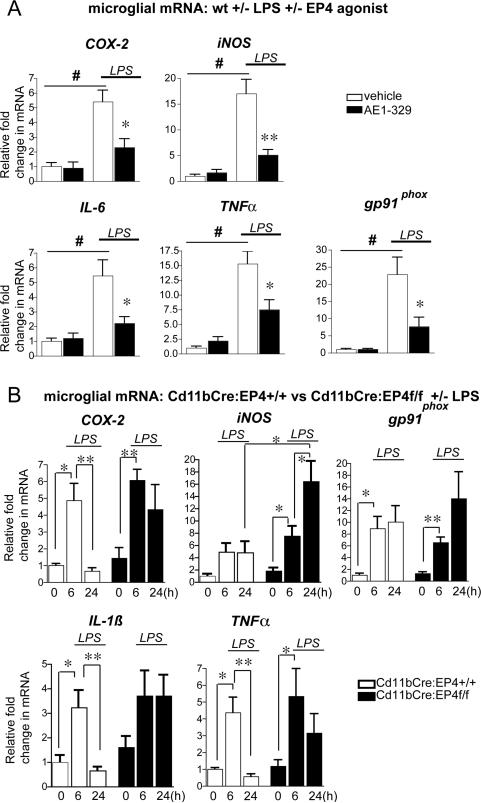
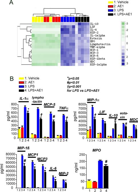
Peripheral inflammation leads to immune responses in brain characterized by microglial activation, elaboration of proinflammatory cytokines and reactive oxygen species, and secondary neuronal injury. The inducible cyclooxygenase (COX), COX-2, mediates a significant component of this response in brain via downstream proinflammatory PG signaling. In this study, we investigated the function of the PGE2 E-prostanoid (EP) 4 receptor in the CNS innate immune response to the bacterial endotoxin LPS. We report that PGE2 EP4 signaling mediates an anti-inflammatory effect in brain by blocking LPS-induced proinflammatory gene expression in mice. This was associated in cultured murine microglial cells with decreased Akt and I-kappaB kinase phosphorylation and decreased nuclear translocation of p65 and p50 NF-kappaB subunits. In vivo, conditional deletion of EP4 in macrophages and microglia increased lipid peroxidation and proinflammatory gene expression in brain and in isolated adult microglia following peripheral LPS administration. Conversely, EP4 selective agonist decreased LPS-induced proinflammatory gene expression in hippocampus and in isolated adult microglia. In plasma, EP4 agonist significantly reduced levels of proinflammatory cytokines and chemokines, indicating that peripheral EP4 activation protects the brain from systemic inflammation. The innate immune response is an important component of disease progression in a number of neurodegenerative disorders, such as Alzheimer's disease, Parkinson's disease, and amyotrophic lateral sclerosis. In addition, recent studies demonstrated adverse vascular effects with chronic administration of COX-2 inhibitors, indicating that specific PG signaling pathways may be protective in vascular function. This study supports an analogous and beneficial effect of PGE2 EP4 receptor signaling in suppressing brain inflammation.


Figures

Figure 1
Figure 1. EP4 receptor expression is dynamically regulated in BV-2 microglial-like cells, in primary microglia, and in hippocampus in response to LPS stimulation
(A) Murine BV-2 cells were stimulated with vehicle or LPS (10ng/ml), and EP4 mRNA was measured at 6h by qPCR (p<0.001; n=3 per condition). (B) EP4 mRNA is also dynamically regulated in rat primary microglia derived from cerebral cortex and hippocampus (ANOVA p<0.001; by post hoc analysis p<0.05 at 1h, p<0.001 at 3h, and p<0.05 at 6h; n=3 per condition). (C) EP4 mRNA is upregulated at 6h in mouse hippocampus and returns to baseline by 24h after peripheral administration of LPS (5 mg/kg IP; n=3-6 per group; p<0.05). (D) Confocal 400X imaging is shown of microglial cells in the hilar region of hippocampus from vehicle-treated and LPS-treated mice (5 mg/kg IP at 6 hours after stimulation). EP4 signal is localized in Iba1 positive microglia (arrows) in a punctate perinuclear distibution in both vehicle and LPS treated mice (scale bar= 10 microns).
Figure 2
Figure 2. EP4 signaling suppresses pro-inflammatory gene transcription in BV-2 cells and primary microglia stimulated with LPS
BV-2 cells (A-D) and cerebral cortical microglia (E) were stimulated with LPS (10 ng/ml) or PBS +/- the EP4 agonist AE1-329 (1 μM) or vehicle. (A) qPCR of COX-2, iNOS, and gp91phox in BV-2 cells at 6h shows a significant increase with LPS treatment in vehicle (v) treated groups (#p<0.001) but a significant decrease with co-administration of AE1-329 (AE; *p<0.05, **p<0.01; n= 3 per condition). (B) Expression in BV-2 cells of pro-inflammatory cytokines TNFα, IL1ß, and IL-6 is significantly induced with LPS (#p<0.001) but decreased with co-stimulation of EP4 receptor agonist (*p<0.05). (C) The anti-inflammatory cytokine IL-10 is upregulated with EP4 receptor stimulation (p<0.05). (D) LPS-induced increase in nitrite concentration in BV-2 cells is decreased in a dose-dependent manner with AE1-329 (0-1μM) at 24 hours (# p<0.001 vehicle vs LPS alone; dose response for AE1-329 ANOVA p<0.0001; post hoc analysis p<0.001 for 0.001, 0.01, 0.1, and 1 μM, n=5 per condition). (E) Primary microglia were stimulated +/- LPS +/- EP4 agonist AE1-329 (100nM) and harvested at 3 hours. qPCR demonstrates a reduced level of iNOS as well as significant reductions of COX-2, TNF-α, and gp91phox and upregulation of IL-10 in LPS-treated microglia with EP4 receptor agonist (#p<0.01-0.001 for vehicle vs LPS; *p<0.05, ***p<0.001 for LPS vs LPS+AE1-329; n=6 per condition).
Figure 3
Figure 3. EP4 receptor activation in BV-2 cells increases PKA activity and reduces LPS-induced phosphorylation of Akt
(A) PKA activity assay of BV-2 cells stimulated with LPS (100 ng/ml), AE1-329 (100 nM), or both shows significant increases with AE1-329 and LPS+AE1-329 (*p<0.05; n=5 samples per condition). (B) Inhibition of PKA with H89 at 5 μM and 10 μM reverses AE1-329-mediated increase in PKA activity (*p<0.05 and **p<0.01). (C) Representative quantitative Western analysis of p-Akt and total Akt shows an increase in p-Akt with LPS (100 ng/ml) treatment that is reduced with stimulation with EP4 agonist AE1-329 (100 nM). BV-2 cells were treated with LPS +/- AE1-329 or vehicle and harvested at time points of 5, 15, 30, and 60 minutes; cell lysates were immunoblotted for phosphorylated Ser473 Akt (p-Akt) and total Akt. The average densitometry from three experiments is shown in the lower panel. p-Akt/Akt values have been normalized to the average signal at time=0 minutes of LPS and LPS+AE1 values. There was a significant effect of AE1-329 treatment [F(1,4)=4.589, p<0.05] and of time [F(1,4)=7.72, p<0.001]. Densitometric measurements of effects of vehicle vs AE1-329 alone did not show differences (data not shown). (D) ELISA of phospho-Thr308 Akt and total Akt at 60 minutes after stimulation with LPS +/- AE1-329 shows a significant increase in p-Akt/Akt levels with LPS stimulation, which is reversed with co-administration of 100 nM AE1-329 (*p<0.05; **p<0.01; n=6 per condition).
Figure 4
Figure 4. EP4 receptor activation in BV-2 cells reduces phosphorylation of IKK and nuclear translocation of NF-κB subunits p65 and p50
(A) Representative quantitative Western analysis of phospho-IKK (p-IKK) and total IKK and densitometric average of three experiments demonstrates an increase in phospho-IKK with LPS stimulation (100 ng/ml) that is significantly attenuated with co-activation of the EP4 receptor (AE1, 100nM; [F(1,4)=4.709, p<0.05] for effect of AE1-329). Densitometric measurements are represented as ratios of p-IKK/IKK and are normalized to time=0 minutes for LPS and LPS+AE1. (B and C) Representative quantitative Western analyses are shown for NF-κB p65 (B) and NF-κB p50 (C) nuclear translocation and cytoplasmic levels in BV-2 cells treated with LPS +/- AE1-329. NF-κB subunit signals were normalized to the nuclear marker lamin B1. Densitometry measurements for nuclear levels of p65 and p50 represent averages of three experiments in which values for individual time points were normalized to the 15 minute vehicle time point. There was a significant effect of AE1-329 treatment for both p65 ([F(1,4)=11.13, p<0.01]) and p50 ([F(1,4)=11.88, p<0.01]) nuclear translocation; there was a significant effect of time for both p65 and p50 [F(1,4)=42.7, p<0.001] and [F(1,4)=27.06, p<0.001], respectively. Maximal attenuation of LPS-dependent nuclear translocation is evident by 60 minutes for NF-κB p65 (**p<0.01) and by 120 minutes for p50 (***p<0.001) with activation of the EP4 receptor. (D) Nuclear translocation of NF-κB p65 was quantified in BV-2 cells 60 minutes after stimulation with either LPS (100 ng/ml) or PBS +/- AE1-329 (100 nM) or vehicle. Cells were immunostained for NF-κB p65 and nuclei were counterstained with Hoechst and examined at 400X with confocal microscopy (scale bar=8 microns). Immunofluorescent staining of p65 in control, AE1-329, and LPS+AE1-329 nuclei (top row in red) demonstrates more diffuse and lighter staining (white horizontal arrows), in contrast to the dense nuclear staining in LPS alone (white vertical arrow). (E) Quantification of immunofluorescent nuclear signal intensity of p65 was carried out in BV-2 cells treated with veh, AE1-329, LPS, and LPS+AE1-329. Five fields per condition were measured, representing >100 cells per field (see Methods). There was a significant increase in nuclear levels of p65 at one hour following LPS stimulation as compared to vehicle alone (***p<0.001), and this increase was significantly attenuated with co-stimulation of the EP4 receptor (**p<0.01).
Figure 5
Figure 5. Cd11bCre conditional deletion of EP4 results in increased pro-inflammatory gene expression and increased lipid peroxidation in brain
Hippocampal mRNA and protein were isolated from Cd11bCre:EP4+/+ and Cd11bCre:EP4f/f male mice 24h after peripheral stimulation with LPS (5mg/kg i.p.). (A) In Cd11bCre:EP4f/f mice qPCR demonstrates increased expression of COX-2, TNFα, IL-6, IL-1ß, and NADPH oxidase subunits p47phox, p67phox, gp91phox, and iNOS 24 hours after peripheral LPS stimulation (* p <0.05; **p<0.01; n = 4–7 male mice per group). (B) Representative quantitative Western analyses and densitometry of p47phox and p67phox in LPS treated Cd11bCre:EP4+/+ versus Cd11bCre:EP4f/f mice (n=4-5 per genotype, *p<0.05, **p<0.01). There were no differences between genotypes treated with vehicle (data not shown). (C) Gas chromatography mass spectrophotometric (GCMS) quantification of lipid peroxidation in cerebral cortical lysates demonstrates a significant increase in F2-isoprostanes (isoPs) in Cd11b: EP4f/f mice 24h after LPS as compared to control Cd11bCre:EP4+/+ mice treated with LPS (n = 4-7 per genotype; *p <0.05).
Figure 6
Figure 6. Systemic administration of EP4 agonist decreases LPS-induced hippocampal pro-inflammatory gene response
Mice were pretreated with AE1-329 (300μg/kg, s.c.) for 30 min before injection of LPS (5mg/kg, i.p.) and hippocampal RNA was isolated at 6 hours after LPS. (A) Pro-inflammatory COX-2 and iNOS are strongly induced 6h after systemic LPS administration, while administration of the selective EP4 agonist AE1-329 blunts induction. (B) Induction of cytokines TNFα, IL-6, and IL-1ß are also decreased with administration of AE1-329 (n=7-8 per group of 3 month male C57B6 mice; # p<0.001 vehicle vs LPS; *p<0.05 LPS/veh vs LPS/AE1-329).
Figure 7
Figure 7. EP4 receptor regulates inflammatory gene expression in microglia isolated from adult mouse brain
Microglia were isolated by density gradient centrifugation from 2-3 mo C57B7 male mice administered saline or LPS +/- EP4 agonist (AE1-329 0.3mg/kg) or vehicle (A), and from 2-3 mo Cd11bCre:EP4+/+ and Cd11bCre:EP4f/f mice 6 hours and 24 hours after LPS (B). (A) Significant increases in microglial expression of COX-2, iNOS, IL-6, TNF-α, and gp91phox were observed in wild type mice in response to LPS, but these increases were significantly blunted with co-treatment with EP4 agonist (#p<0.01; (*p<0.05; **p<0.01; n=6-8 mice per group). (B) Proinflammatory gene expression is elevated in Cd11bCre:EP4+/+ and Cd11bCre:EP4f/f microglia at 6 hours after LPS; however, increased gene expression persists at 24h in microglia isolated from Cd11bCre:EP4f/f mice as compared to Cd11bCre:EP4+/+ control mice. Gene expression does not return to basal levels for COX-2, IL-1ß, and TNF-α at 24h in Cd11bCre:EP4f/f microglia, and is significantly increased beyond the 6 hour level for iNOS at this late time point (*p<0.05; **p<0.01; n=4-8 per group).
Figure 8
Figure 8. Peripheral administration of EP4 reduces LPS-mediated inflammatory response in plasma
Plasma was collected and analyzed at 3 hours after co-administration of PBS or LPS (5mg/kg, i.p.) +/- vehicle or AE1-329 (300μg/kg, s.c.). (A) Cluster analysis of regulated cytokines and myeloperoxidase (MPO) following peripheral PBS or LPS administration +/- AE1-329. (B) Absolute concentrations of regulated cytokines (pg/ml) and MPO (ng/ml) are decreased with AE1-329 administration.

Similar articles

Cited by

References

    1. Nguyen MD, Julien JP, Rivest S. Innate immunity: the missing link in neuroprotection and neurodegeneration? Nature reviews. 2002;3:216–227. - PubMed
    1. Fassbender K, Walter S, Kuhl S, Landmann R, Ishii K, Bertsch T, Stalder AK, Muehlhauser F, Liu Y, Ulmer AJ, Rivest S, Lentschat A, Gulbins E, Jucker M, Staufenbiel M, Brechtel K, Walter J, Multhaup G, Penke B, Adachi Y, Hartmann T, Beyreuther K. The LPS receptor (CD14) links innate immunity with Alzheimer's disease. Faseb J. 2004;18:203–205. - PubMed
    1. Walter S, Letiembre M, Liu Y, Heine H, Penke B, Hao W, Bode B, Manietta N, Walter J, Schulz-Schuffer W, Fassbender K. Role of the toll-like receptor 4 in neuroinflammation in Alzheimer's disease. Cell Physiol Biochem. 2007;20:947–956. - PubMed
    1. Moisse K, Strong MJ. Innate immunity in amyotrophic lateral sclerosis. Biochim Biophys Acta. 2006;1762:1083–1093. - PubMed
    1. Letiembre M, Liu Y, Walter S, Hao W, Pfander T, Wrede A, Schulz-Schaeffer W, Fassbender K. Screening of innate immune receptors in neurodegenerative diseases: A similar pattern. Neurobiol Aging. 2007 - PubMed

Publication types

MeSH terms

Substances