. 2010 Jun 1;184(11):6199-208.
doi: 10.4049/jimmunol.1000078. Epub 2010 May 5.

HLA-F complex without peptide binds to MHC class I protein in the open conformer form

Affiliations

HLA-F complex without peptide binds to MHC class I protein in the open conformer form

Jodie P Goodridge et al. J Immunol. .

Abstract

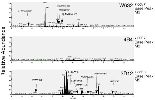
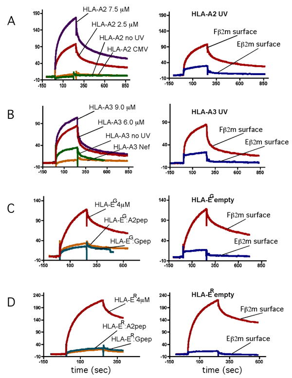
HLA-F has low levels of polymorphism in humans and is highly conserved among primates, suggesting a conserved function in the immune response. In this study, we probed the structure of HLA-F on the surface of B lymphoblastoid cell lines and activated lymphocytes by direct measurement of peptide binding to native HLA-F. Our findings suggested that HLA-F is expressed independently of bound peptide, at least in regard to peptide complexity profiles similar to those of either HLA-E or classical MHC class I (MHC-I). As a further probe of native HLA-F structure, we used a number of complementary approaches to explore the interactions of HLA-F with other molecules, at the cell surface, intracellularly, and in direct physical biochemical measurements. This analysis demonstrated that HLA-F surface expression was coincident with MHC-I H chain (HC) expression and was downregulated upon perturbation of MHC-I HC structure. It was further possible to directly demonstrate that MHC-I would interact with HLA-F only when in the form of an open conformer free of peptide and not as a trimeric complex. This interaction was directly observed by coimmunoprecipitation and by surface plasmon resonance and indirectly on the surface of cells through coincident tetramer and MHC-I HC colocalization. These data suggest that HLA-F is expressed independently of peptide and that a physical interaction specific to MHC-I HC plays a role in the function of MHC-I HC expression in activated lymphocytes.


Figures

Figure 1
Figure 1
Peptide elution from MHC class I. MS elution times are charted against relative abundance for each of three MHC class I antibodies W6/32 (pan class I), 4B4 (anti-HLA-F), and 3D12 (anti-HLA-E) as indicated. The relative intensity of the highest peak within each profile is indicated to the right of each chromatogram. The positions and sequences of peptides that had confirmed sequences via MS/MS are indicated within each profile. None of the putative peptides in the 4B4 profile were found in more than one run, and it was not possible to confirm any via MS/MS.
Figure 2
Figure 2
HLA-F surface expression is downregulated in response to MHC-I heavy chain mAb. (A) B-LCL SAVC cells without (solid lines) and following addition of MHC-I heavy chain mAbs HCA2, LA45, or anti-HLA-F 3D11 as indicated within brackets (dotted lines). The reagent used for staining in each FACS profile is indicated immediately above the bracketed mAb with control mAb (grey). (B) Differential downregulation of HLA-F in response to mAb HCA2 and LA45 in B-LCL cell lines with and without reactive MHC class I. Cell line names and reactivities with the respective mAb are indicated directly beneath each set of bars. Anti-F mAb 3D11 reactivity is plotted after the addition of HCA2, LA45, and W6/32 with bars according to the key on the right. (C) Relative staining of anti-F mAb 3D11 on B-LCL AMAI cells without HCA2 (solid lines) and following HCA2 addition (dotted lines) with control mAb (grey). Cells were untreated (left) and treated with 100uM NEM (right) as indicated.
Figure 3
Figure 3
Acid induced free class I heavy chain expression facilitates HLA-F tetramer binding. HLA-F negative and free Class I Heavy Chain negative T cell lines Jurkat, Molt-3, HUT78, H9 and monocytic cell line U937, as well as Class I bare cell line Daudi were acid treated and subsequently stained for free Class I heavy chain (HCA2) and for HLA-F tetramer binding. (A) HCA2 reactivity (solid line) does not increase upon acid treatment (dashed line) for class I bare cell line Daudi, while T cell line H9 becomes both HCA2 positive (left panel, dashed line) and HLA-F tetramer reactive (right panel, dashed line) after acid treatment. (B) Comparison of HCA2 reactivity with HLA-F tetramer staining in the 6 cell lines upon acid treatment reveals a strong association between free class I heavy chain expression and HLA-F tetramer binding. Shaded diamonds designate cells before acid treatment and open diamonds after acid treatment. (C) Cell line OSP2 examined before (solid line) and after (dotted line) CD3 + CD28 mAb treatment and examined for MHCI-HC expression and F tetramer binding as indicated. (D) Immunofluorescent staining of CTL clone 1C7-7 which expresses levels of HLA-F and HCA2 reactive MHC-I HC, and H9 cells after acid treatment (from A) stained with HLA-F tetramer and HCA2 individually as indicated with overlapping profiles at the right.
Figure 4
Figure 4
MHCI-HC, including HLA-E HC, co-precipitate with HLA-F. Cells were lysed directly in NP40 buffer containing freshly prepared 50 mM iodoacetamide. Three sets of sequential immunoprecipitations of total cell lysate from LCL PLH were carried out and separated on a reducing 10% T-G gel. Each set of sequential IPs were carried out starting and finishing with different mAbs of the group of three as indicated above each lane. The first serial set included in the leftmost three lanes of each gel started with IP using 4B4 and loaded in lane 1, after which the remaining material was subjected to IP with 3D11 and run in lane 2, and again the remaining lysate subjected to IP with 4A11 and run in lane 3. Two additional serial IPs were carried out and run in lanes 4-6 and 7-9 using mAbs as indicated above each lane and grouped together with bars immediately below each mAb designation. After fractionation each gel was blotted and analyzed via western analysis as described in Methods using the mAb for detection indicated immediately beneath each image. All three gels shown were identically constructed using the order of sequential IPs as indicated above each lane. The positions of molecular weight markers are indicated to the left and right.
Figure 5
Figure 5
MHC class I heavy chain, but not complex, binds directly and specifically to HLA-F. Surface plasmon resonance binding sensograms were analyzed using surfaces coated with refolded F β2m on the left or with two channels simultaneously, containing Fβ2m and EGβ2m separately, shown in the rightmost panels. (A) HLA-A2 refolded with conditional ligand as described in Materials and Methods was injected over the Fβ2m surface after UV exposure at 2.5 μM (red line) and 7.5 μM (purple line), and HLA-A2 refolded with conditional ligand with no UV at 5.0 μM (green line) and refolded with CMV pp65 (495-503) peptide at 4μM (orange line) all as indicated. HLA-A2 refolded with conditional ligand and UV treated was passed over the separate and marked Fβ2m (red line) and EGβ2m (blue line) surfaces simultaneously in the panel immediately to the right. (B) HLA-A3 refolded with conditional ligand was injected after UV exposure at 6 μM (red line) and 9.5 μM (purple line) and HLA-A3 refolded with conditional ligand with no UV at 14 μM (green line) and refolded with HIV Nef (73-82) peptide at 10μM (orange line) all as indicated. HLA-A3 refolded with conditional ligand and UV treated was passed over the Fβ2m (red line) and EGβ2m (blue line) surfaces simultaneously as indicated. (C) HLA-EG + β2m refolded without peptide at 4μM (purple line) and refolded with nonamer from HLA-A2 (orange line) and HLA-G (green line) were injected over the Fβ2m surface. HLA-EG + β2m refolded without peptide at 4μM was recorded over the Fβ2m (red line) and EGβ2m (blue line) surfaces simultaneously as indicated. (D) Identical to (C) using refolded HLA-ER heavy chain.

Similar articles

Cited by

References

    1. Accolla RS, Adorini L, Sartoris S, Sinigaglia F, Guardiola J. MHC: orchestrating the immune response. Immunol Today. 1995;16:8–11. - PubMed
    1. Bauer S, Groh V, Wu J, Steinle A, Phillips JH, Lanier LL, Spies T. Activation of NK cells and T cells by NKG2D, a receptor for stress-inducible MICA. Science. 1999;285:727–729. - PubMed
    1. Gonzalez S, Groh V, Spies T. Immunobiology of human NKG2D and its ligands. Curr Top Microbiol Immunol. 2006;298:121–138. - PubMed
    1. Lawlor DA, Zemmour J, Ennis PD, Parham P. Evolution of class-I MHC genes and proteins: from natural selection to thymic selection. Annu Rev Immunol. 1990;8:23–63. - PubMed
    1. Pamer E, Cresswell P. Mechanisms of MHC class I--restricted antigen processing. Annu Rev Immunol. 1998;16:323–358. - PubMed

Publication types