. 2010 Jun 1;184(11):6209-22.
doi: 10.4049/jimmunol.1000231. Epub 2010 May 5.

Vgamma2Vdelta2 T Cell Receptor recognition of prenyl pyrophosphates is dependent on all CDRs

Affiliations

Vgamma2Vdelta2 T Cell Receptor recognition of prenyl pyrophosphates is dependent on all CDRs

Hong Wang et al. J Immunol. .

Abstract

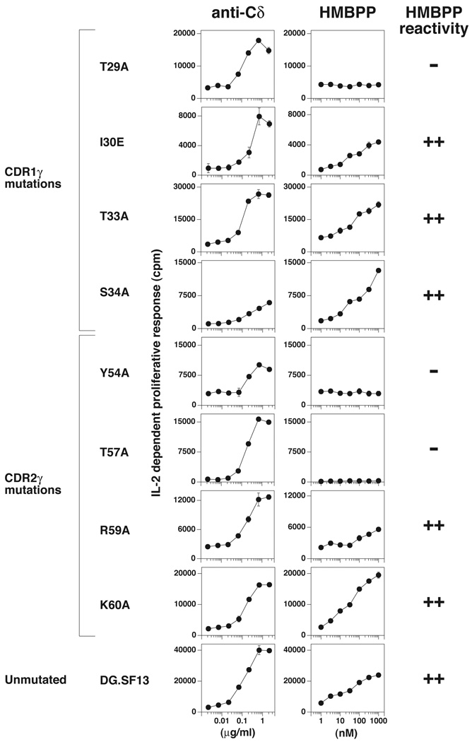
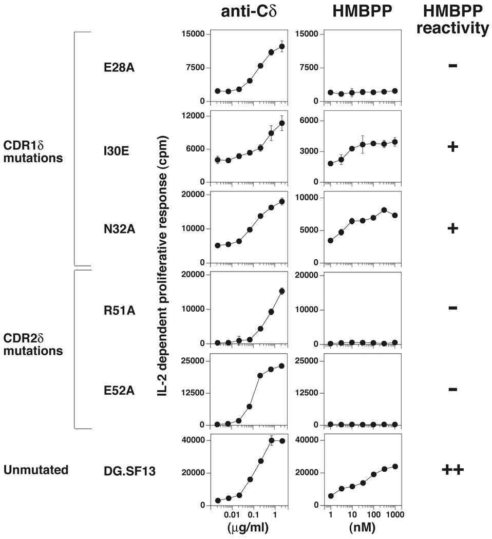
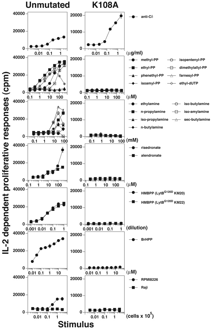
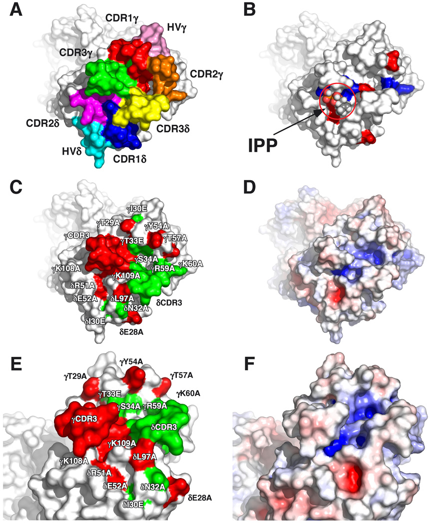
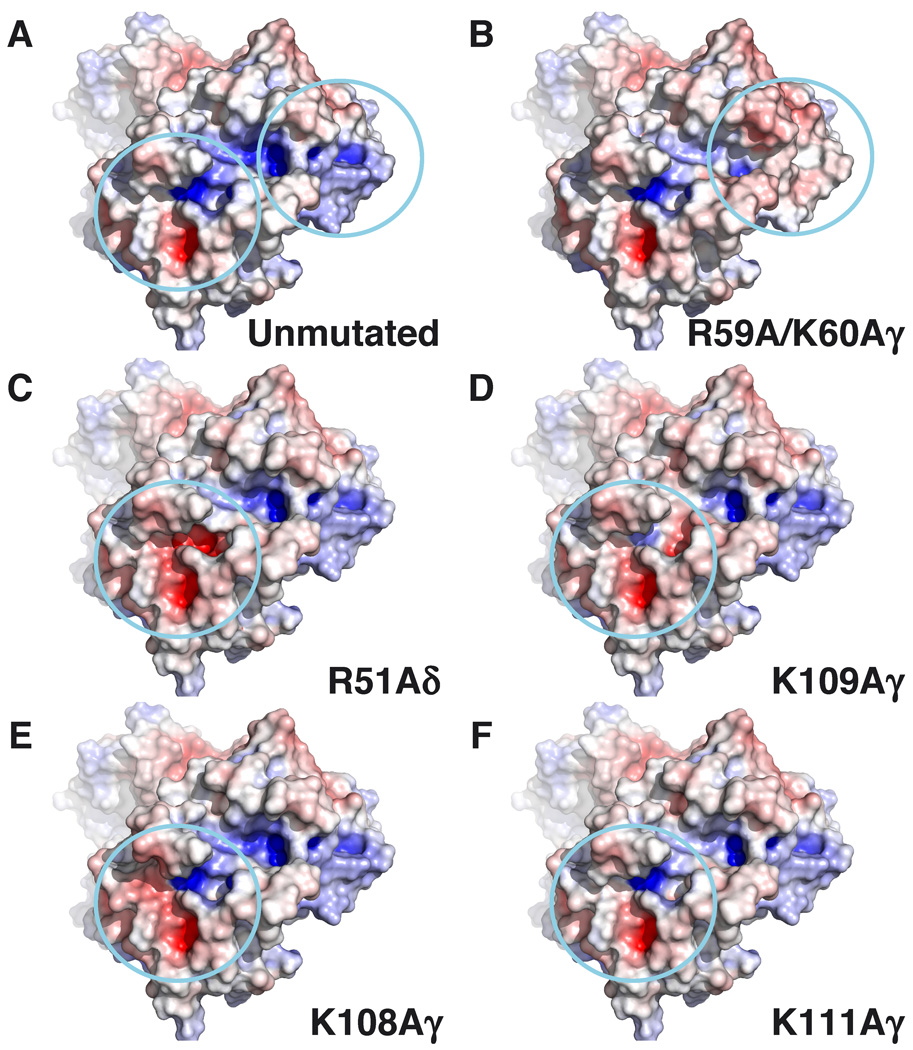
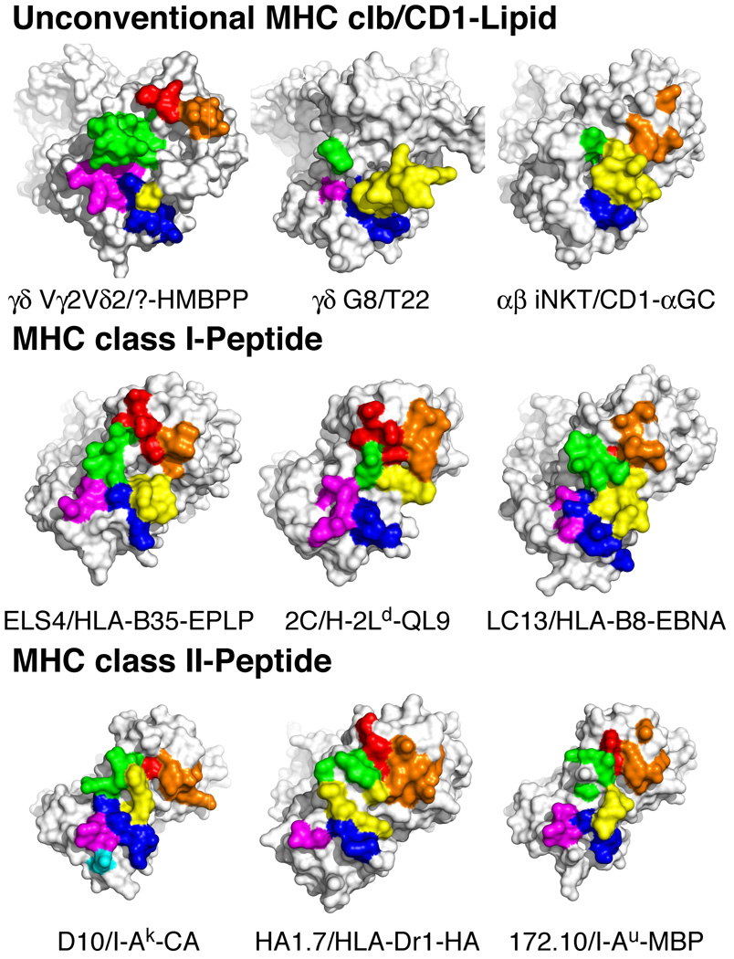
gammadelta T cells differ from alphabeta T cells in the Ags they recognize and their functions in immunity. Although most alphabeta TCRs recognize peptides presented by MHC class I or II, human gammadelta T cells expressing Vgamma2Vdelta2 TCRs recognize nonpeptide prenyl pyrophosphates. To define the molecular basis for this recognition, the effect of mutations in the TCR CDR was assessed. Mutations in all CDR loops altered recognition and cover a large footprint. Unlike murine gammadelta TCR recognition of the MHC class Ib T22 protein, there was no CDR3delta motif required for recognition because only one residue is required. Instead, the length and sequence of CDR3gamma was key. Although a prenyl pyrophosphate-binding site was defined by Lys109 in Jgamma1.2 and Arg51 in CDR2delta, the area outlined by critical mutations is much larger. These results show that prenyl pyrophosphate recognition is primarily by germline-encoded regions of the gammadelta TCR, allowing a high proportion of Vgamma2Vdelta2 TCRs to respond. This underscores its parallels to innate immune receptors. Our results also provide strong evidence for the existence of an Ag-presenting molecule for prenyl pyrophosphates.


Conflict of interest statement

Disclosures

The authors have no conflicting financial interests.

Figures

FIGURE 1
FIGURE 1. Recognition of HMBPP by Vγ2Vδ2 TCR transfectants expressing mutant Vγ2 chains
J.RT3-T3.5 β Jurkat cells were transfected with unmutated or mutated DG.SF13 TCR-γ cDNAs together with the unmutated DG.SF13 TCR-δ chain cDNA. After drug selection, anti-Cδ responsive transfectants were identified and stimulated with HMBPP in the presence of Va2 APCs and 2.5 ng/ml PMA. The anti-Cδ mAb (anti-TCRδ1) and HMBPP were added to the cultures stating at 2.15 µg/ml and 1000 nM, respectively, and serially diluted by half-log intervals. After 24 h, the culture supernatants were harvested and assayed for IL-2 activity using the IL-2-dependent cell line, HT2. Results from one transfectant are shown for each mutation and are representative of the results obtained with two to four other independently derived transfectants (Supplemental Fig. 1). Values shown are mean ± SEM of duplicate or triplicate samples. HMBPP reactivity was considered (++) if the maximum HMBPP response was >40% of the control anti-Cδ response, (+) if between 20–40% of the control response, and (−) if <10% of the control response.
FIGURE 2
FIGURE 2. Recognition of HMBPP by Vγ2Vδ2 TCR transfectants expressing mutant Vδ2 chains
J.RT3-T3.5 β Jurkat cells were transfected with unmutated or mutated DG.SF13 TCR-δ cDNAs together with the unmutated DG.SF13 TCR-γ chain cDNA. Culture conditions and the IL-2 assay were as in Fig. 1. Results from one transfectant are shown for each mutation and are representative of the results obtained with one to four other independently derived transfectants (Supplemental Fig. 2).
FIGURE 3
FIGURE 3. Critical role of K108 in the CDR3 of Vγ2 in the recognition of nonpeptide Ags and tumor cells
J.RT3-T3.5 β Jurkat cells were transfected with the DG.SF13 TCR-γ chain cDNA with a lysine (K) to alanine (A) mutation at position 108 in the CDR3γ region and the unmutated DG.SF13 TCR-δ cDNA. The transfectant obtained was named K108A and compared with an unmutated DG.SF13 TCR transfectant for stimulation by anti-Cδ, prenyl pyrophosphates, alkylamines, bisphosphonates (risedronate and alendronate), HMBPP (in supernatants of the E.coli lytB mutants, KM20, and KM22), bromohydrin pyrophosphate, and lymphoma cell lines (RPMI 8226 and Raji). Culture conditions and the IL-2 assay were as in Fig. 1.
FIGURE 4
FIGURE 4. Recognition of prenyl pyrophosphates by human Vγ2Vδ2 TCR is dependent on all CDRs
A, Top-down view of the Vγ2Vδ2 TCR with complementarity determining regions for the γ and δ chains labeled. CDR residues are colored such that those in: CDR1δ are blue, CDR1δ are magenta, CDR3δ are yellow, HV4δ are cyan, CDR1γ are red, CDR2γ are orange, CDR3γ are green, and HV4γ are pink. B, Top-down view of the Vγ2Vδ2 TCR with basic amino acids colored blue and acidic amino acids colored red. IPP is shown in the potential prenyl pyrophosphate-binding site for size comparison. C, Top-down view of critical amino acid residues that disrupt prenyl pyrophosphate recognition are colored red on the Vγ2Vδ2 TCR whereas non-critical amino acid residues are colored green. D, Top-down view of the surface potential of the Vγ2Vδ2 TCR (colored from red [−8 kT] to blue [+8 kT]) reveals two positively charged potential binding sites. E, Side view of critical amino acid residues that disrupt prenyl pyrophosphate recognition are colored red on the Vγ2Vδ2 TCR whereas non-critical amino acid residues are colored green. F, Side view of the surface potential of the Vγ2Vδ2 TCR (colored as in D).
FIGURE 5
FIGURE 5. Surface potential effect of in silico alanine mutations of basic residues in the Vγ2Vδ2 TCR
Basic residues were mutated to alanine in silico using Pymol (DeLano Scientific), and the surface potential of the mutated TCRs were calculated using the APBS plugin in Pymol. The TCRs are oriented with the γ chains on the top left and the δ chains on the bottom right of the panels. Top down views of the surface potential of the Vγ2Vδ2 TCR are shown (colored from red (−8 kT) to blue (+8 kT). The two positively charged regions are circled. A, Surface potential for the unmutated Vγ2Vδ2 TCR. B–F, Effect on surface potential of alanine replacement of R59A and K60Aγ (B), R51Aδ (C), K109Aγ (D), K108Aγ (E), and K111Aγ (F).
FIGURE 6
FIGURE 6. Potential contact residues of the Vγ2Vδ2 TCR for a putative antigen presenting molecule-prenyl pyrophosphate complex differs from other unconventional TCRs and from conventional TCRs specific for MHC-peptide complexes
The TCRs are oriented with the β/γ chains on the top left and the α/δ chains on the bottom right of the panels. Contact residues are colored such that those in: CDR1α/δ are blue, CDR2α/δ are magenta, CDR3α/δ are yellow, HV4α/δ are cyan, CDR1β/γ are red, CDR2β/γ are orange, and CDR3β/γ are green. The diagonal orientation does not attempt to match the docking angle on the MHC/CD1 ligand. Top panels, Putative contact residue "footprint" of the Vγ2Vδ2 TCR based on the effects of point mutations on human Vγ2Vδ2 T cell reactivity to nonpeptide Ags (top left panel). Note the potential large contribution of germline-encoded regions of CDR3γ compared with only a minor contribution by L97 in CDR3δ. In contrast, the G8 γδ TCR (top middle panel), that is specific for the T22 MHC class Ib molecule, predominantly uses CDR3δ for recognition. The iNKT αβ TCR (top left panel) binding to the CD1d-α-GalCer complex also predominantly uses the CDR3α region (which corresponds to CDR3δ). Middle panels, Conventional αβ TCRs specific for MHC class I-foreign peptide complexes generally use both CDR3α and CDR3β for MHC-peptide recognition in addition to CDR1 and CDR2. Bottom panels, Conventional αβ TCRs specific for MHC class II/peptide complexes are similar to MHC class I/peptide-specific αβ TCRs and involve extensive CDR3α and CDR3β contacts in addition to CDR1 and CDR2 contacts. Additional examples of αβ TCR recognition of MHC-peptide complexes are shown in Supplemental Figs. 4 and 5.

Similar articles

Cited by

References

    1. Gapin L. Where do MAIT cells fit in the family of unconventional T cells? PLoS Biol. 2009;7:435–438. - PMC - PubMed
    1. Cerundolo V, Silk JD, Masri SH, Salio M. Harnessing invariant NKT cells in vaccination strategies. Nat. Rev. Immunol. 2009;9:28–38. - PubMed
    1. Kronenberg M, Engel I. On the road: progress in finding the unique pathway of invariant NKT cell differentiation. Curr. Opin. Immunol. 2007;19:186–193. - PubMed
    1. Morita CT, Parker CM, Brenner MB, Band H. T cell receptor usage and functional capabilities of human γδ T cells at birth. J. Immunol. 1994;153:3979–3988. - PubMed
    1. Parker CM, Groh V, Band H, Porcelli SA, Morita C, Fabbi M, Glass D, Strominger JL, Brenner MB. Evidence for extrathymic changes in the T cell receptor γ/δ repertoire. J. Exp. Med. 1990;171:1597–1612. - PMC - PubMed

Publication types

MeSH terms

Substances