. 2010 Jun 1;184(11):6438-46.
doi: 10.4049/jimmunol.0902009. Epub 2010 May 5.

Arginase I suppresses IL-12/IL-23p40-driven intestinal inflammation during acute schistosomiasis

Affiliations

Arginase I suppresses IL-12/IL-23p40-driven intestinal inflammation during acute schistosomiasis

De'Broski R Herbert et al. J Immunol. .

Abstract

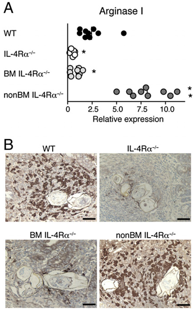
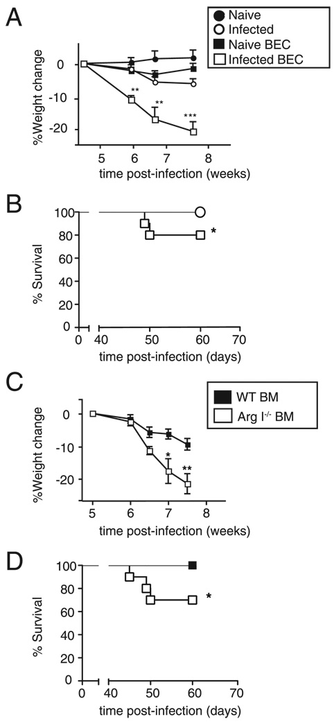
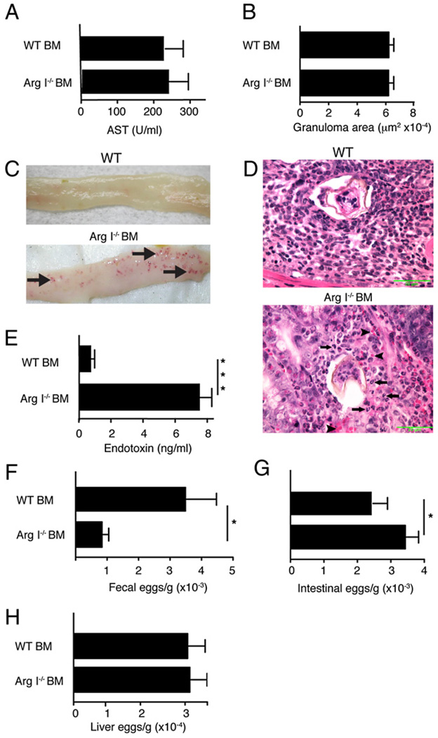
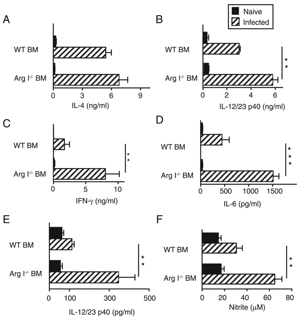
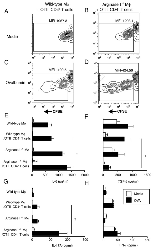
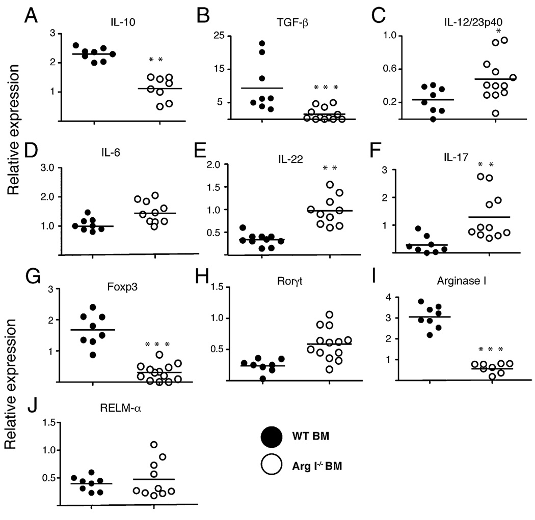
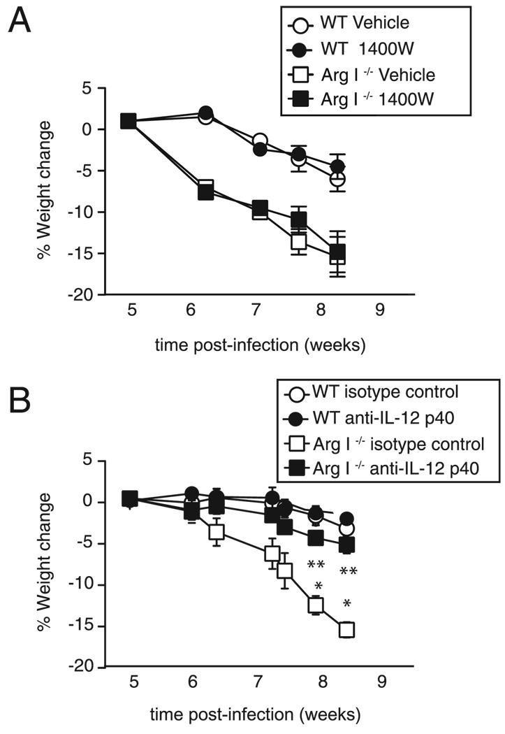
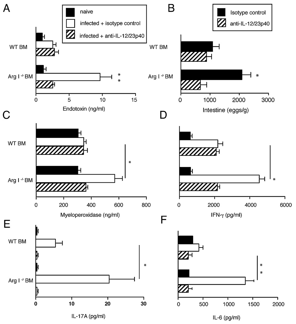
Alternatively activated macrophages prevent lethal intestinal pathology caused by worm ova in mice infected with the human parasite Schistosoma mansoni through mechanisms that are currently unclear. This study demonstrates that arginase I (Arg I), a major product of IL-4- and IL-13-induced alternatively activated macrophages, prevents cachexia, neutrophilia, and endotoxemia during acute schistosomiasis. Specifically, Arg I-positive macrophages promote TGF-beta production and Foxp3 expression, suppress Ag-specific T cell proliferation, and limit Th17 differentiation. S. mansoni-infected Arg I-deficient bone marrow chimeras develop a marked accumulation of worm ova within the ileum but impaired fecal egg excretion compared with infected wild-type bone marrow chimeras. Worm ova accumulation in the intestines of Arg I-deficient bone marrow chimeras was associated with intestinal hemorrhage and production of molecules associated with classical macrophage activation (increased production of IL-6, NO, and IL-12/IL-23p40), but whereas inhibition of NO synthase-2 has marginal effects, IL-12/IL-23p40 neutralization abrogates both cachexia and intestinal inflammation and reduces the number of ova within the gut. Thus, macrophage-derived Arg I protects hosts against excessive tissue injury caused by worm eggs during acute schistosomiasis by suppressing IL-12/IL-23p40 production and maintaining the Treg/Th17 balance within the intestinal mucosa.


Conflict of interest statement

Disclosures

The authors have no financial conflicts of interest.

Figures

FIGURE 1
FIGURE 1
Arg I production in S. mansoni granulomas requires IL-4Rα expression only on BM-derived cells. A, Intestinal mRNA transcripts for Arg I were quantitated by real-time PCR 7.4 wk postinoculation in S. mansoni-infected BM chimeras that expressed IL-4Rα on all cells (WT), lacked IL-4Rα expression on all cells (IL-4Rα−/−), lacked IL-4Rα expression on BM-derived cells (BMIL-4Rα−/−), or lacked IL-4Rα expression on non–BM-derived cells (non–BMIL-4Rα−/−). Transcript levels are expressed as fold increase compared with naive WT tissue. Experiment performed three times with similar results. n = 6–8 mice/group; *p < 0.05; **p < 0.01 compared with WT. B, Immunohistochemical staining for Arg I was performed 7.4 wk postinfection on intestinal granulomas from WT (top left panel), IL-4Rα−/− (top right panel), BMIL-4Rα−/− (bottom left panel), and BMIL-4Rα−/− (bottom right panel) mice. Original magnification ×400. Representative photos from 150 granulomas examined per group. Positive staining indicated by dark brown color.
FIGURE 2
FIGURE 2
Mice treated with an arginase antagonist or chimeras lacking BM-derived Arg I develop cachexia and increased mortality during acute schistosomiasis. Weight change (A) and survival (B) of naive or S. mansoni-infected mice that were administered normal drinking water or drinking water containing BEC. Weight change (C) and survival (D) of WT and Arg I−/− BM chimeras following inoculation with 60–70 S. mansoni cercariae. Representative of three independent experiments with 8–10 mice/group. *p < 0.05; **p < 0.01; ***p < 0.001 compared with WT-infected group.
FIGURE 3
FIGURE 3
BM Arg I deficiency causes increased tissue injury, ova accumulation, and neutrophilic inflammation in the intestine during acute schistosomiasis. Serum levels of AST (A) and liver granuloma size (B). Representative of 150 granulomas evaluated with 8–10 mice/group. C, Representative photographs of terminal ileum from S. mansoni-infected WT BM or Arg I−/− BM chimeras 7.4 wk postinoculation. Note areas of hemorrhage (arrows). D, Representative photomicrographs of intestinal granulomas in WT BM and Arg I−/− BM chimeras at 7.5 wk postworm inoculation. Arrowheads point to erythrocytes, and arrows indicate neutrophils. Fifty granulomas examined per group. Original magnification ×600. Serum endotoxin levels (E), number of parasite ova in feces (F), intestine (G), and liver (H) at 7.4 wk postinoculation. Representative of three independent experiments with 8–10 mice/group. *p < 0.05; **p < 0.01; ***p < 0.001 compared with WT-infected group. AST, aspartate aminotransferase.
FIGURE 4
FIGURE 4
Arg I-deficient BM chimeras produce elevated proinflammatory cytokines and show evidence of classical Mφ activation. Systemic production of IL-4 (A), IL-12/IL-23p40 (B), IFN-γ (C), and IL-6 (D) in WT and Arg I−/− BM chimeras analyzed 7.4 wk postinoculation. Adherent peritoneal cells from S. mansoni-infected WT BM or Arg I−/− BM chimeras were stimulated with 1 µg/ml Escherichia coli endotoxin for 24 h and supernatants analyzed for levels of IL-12/IL-23p40 (E) and nitrite (F). Representative of three independent experiments with mean ± SE, n = 8–10 mice/group. *p < 0.05; **p < 0.01.
FIGURE 5
FIGURE 5
Arg I-deficient macrophages drive Ag-specific CD4+ T cell proliferation and preferentially induce Th17 differentiation. Contour plots showing MFI of CFSE within OTII CD4+ T cells following coculture with (A, C) WT or (B, D) Arg I-deficient macrophages that were either (A, B) untreated or (C, D) pulsed with 50 µg/ml OVA. Analysis was performed at 96 h. From the experiment shown above, supernatants were analyzed for production of IL-6 (E), TGF-β (F), IL-17 (G), and IFN-γ (H) by ELISA. □ media alone; ■, OVA. Representative of three independent experiments with mean ± SE of triplicate wells. *p < 0.05; **p < 0.01. MFI, mean fluorescence intensity.
FIGURE 6
FIGURE 6
Arg I-deficient BM chimeras show decreased expression of immunosuppressive cytokines and increased Th17-associated cytokines in the intestine during acute schistosomiasis. Intestinal mRNA levels for murine IL-10 (A), TGF-β (B), IL-12/IL-23p40 (C), IL-6 (D), IL-17 (E), IL-22 (F), Foxp3 (G), Rorγt (H), ArgI (I), and RELMα (J), quantitated by real-time PCR 7.4 wk post-S. mansoni inoculation of BM chimeras. Representative of three independent experiments with mean ± SE, n = 8–10 mice/group. *p < 0.05; **p < 0.01.
FIGURE 7
FIGURE 7
IL-12/IL-23p40 neutralization but not NOS-2 inhibition rescues Arg I−/− BM chimeras from cachexia. Weight change of S. mansoni-infected WT and Arg I−/− BM chimeras that were administered 2 mg biweekly injections of the NOS-2 antagonist 1400W versus saline (vehicle) (A) or 2 mg rat anti–IL-12/IL-23p40 (C17.8) versus rat IgG2a (GL117) isotype control mAb (B) starting 5 wk postworm inoculation. Representative of two independent experiments n = 8–10 mice/group. *p < 0.05; **p < 0.01.
FIGURE 8
FIGURE 8
IL-12/IL-23p40 neutralization abrogates intestinal inflammation and injury and reduces S. mansoni intestinal egg accumulation in Arg I−/− BM chimeras. Levels of serum endotoxin (A), number of parasite ova in intestine (B), serum MPO (C), IFN-γ (D), IL-17A (E), and IL-6 (F) produced from ileal punch biopsies and serum levels of IFN-γ and IL-17A analyzed 8 wk postworm inoculation. Mean ± SE of 8–10 mice/group. Representative of two independent experiments. *p < 0.05; **p < 0.01.

References

    1. Urban JF, Jr, Noben-Trauth N, Schopf L, Madden KB, Finkelman FD. Cutting edge: IL-4 receptor expression by non-bone marrow-derived cells is required to expel gastrointestinal nematode parasites. J. Immunol. 2001;167:6078–6081. - PubMed
    1. Chitsulo L, Loverde P, Engels D. Schistosomiasis. Nat. Rev. Microbiol. 2004;2:12–13. - PubMed
    1. Gryseels B, Polman K, Clerinx J, Kestens L. Human schistosemiasis. Lancet. 2006;368:1106–1118. - PubMed
    1. Herbert DR, Orekov T, Perkins C, Finkelman FD. IL-10 and TGF-β redundantly protect against severe liver injury and mortality during acute schistosomiasis. J. Immunol. 2008;181:7214–7220. - PMC - PubMed
    1. La Flamme AC, Patton EA, Bauman B, Pearce EJ. IL-4 plays a crucial role in regulating oxidative damage in the liver during schistosomiasis. J. Immunol. 2001;166:1903–1911. - PubMed

Publication types

MeSH terms