

Epidermal T cells and wound healing
- PMID: 20483798
- PMCID: PMC2944652
- DOI: 10.4049/jimmunol.0902733
Epidermal T cells and wound healing
Abstract
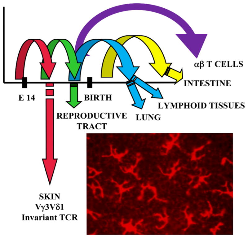
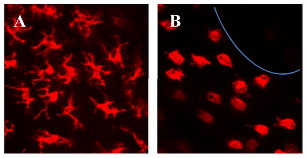
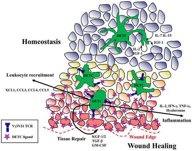
The murine epidermis contains resident T cells that express a canonical gammadelta TCR. These cells arise from fetal thymic precursors and use a TCR that is restricted to the skin in adult animals. These cells assume a dendritic morphology in normal skin and constitutively produce low levels of cytokines that contribute to epidermal homeostasis. When skin is wounded, an unknown Ag is expressed on damaged keratinocytes. Neighboring gammadelta T cells then round up and contribute to wound healing by local production of epithelial growth factors and inflammatory cytokines. In the absence of skin gammadelta T cells, wound healing is impaired. Similarly, epidermal T cells from patients with healing wounds are activated and secreting growth factors. Patients with nonhealing wounds have a defective epidermal T cell response. Information gained on the role of epidermal-resident T cells in the mouse may provide information for development of new therapeutic approaches to wound healing.
Figures
References
-
- Hayday A, Viney JL. The ins and outs of body surface immunology. Science. 2000;290:97–100. - PubMed
-
- Havran WL, Jameson JM, Witherden DA. Epithelial cells and their neighbors. III. Interactions between intraepithelial lymphocytes and neighboring epithelial cells. Am J Physiol Gastrointest Liver Physiol. 2005;289:G627–630. - PubMed
-
- Hayday AC. γδ T cells and the lymphoid stress-surveillance response. Immunity. 2009;31:184–196. - PubMed
-
- Jameson J, Havran WL. Skin γδ T-cell functions in homeostasis and wound healing. Immunol Rev. 2007;215:114–122. - PubMed
Publication types
MeSH terms
Substances
Grants and funding
LinkOut - more resources
Full Text Sources
Other Literature Sources
