

Sarcolemmal nNOS anchoring reveals a qualitative difference between dystrophin and utrophin
- PMID: 20483958
- PMCID: PMC2880012
- DOI: 10.1242/jcs.064808
Sarcolemmal nNOS anchoring reveals a qualitative difference between dystrophin and utrophin
Abstract
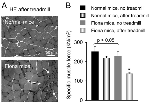
Duchenne muscular dystrophy (DMD) is a lethal muscle disease caused by dystrophin deficiency. In normal muscle, dystrophin helps maintain sarcolemmal stability. Dystrophin also recruits neuronal nitric oxide synthase (nNOS) to the sarcolemma. Failure to anchor nNOS to the membrane leads to functional ischemia and aggravates muscle disease in DMD. Over the past two decades, a great variety of therapeutic modalities have been explored to treat DMD. A particularly attractive approach is to increase utrophin expression. Utrophin shares considerable sequence, structural and functional similarity with dystrophin. Here, we test the hypothesis that utrophin also brings nNOS to the sarcolemma. Full-length utrophin cDNA was expressed in dystrophin-deficient mdx mice by gutted adenovirus or via transgenic overexpression. Subcellular nNOS localization was determined by immunofluorescence staining, in situ nNOS activity staining and microsomal preparation western blot. Despite supra-physiological utrophin expression, we did not detect nNOS at the sarcolemma. Furthermore, transgenic utrophin overexpression failed to protect mdx muscle from exercise-associated injury. Our results suggest that full-length utrophin cannot anchor nNOS to the sarcolemma. This finding might have important implications for the development of utrophin-based DMD therapies.
Figures


Similar articles
-
Dystrophin R16/17 protein therapy restores sarcolemmal nNOS in trans and improves muscle perfusion and function.Mol Med. 2019 Jul 2;25(1):31. doi: 10.1186/s10020-019-0101-6. Mol Med. 2019. PMID: 31266455 Free PMC article.
-
Dystrophins carrying spectrin-like repeats 16 and 17 anchor nNOS to the sarcolemma and enhance exercise performance in a mouse model of muscular dystrophy.J Clin Invest. 2009 Mar;119(3):624-35. doi: 10.1172/JCI36612. Epub 2009 Feb 23. J Clin Invest. 2009. PMID: 19229108 Free PMC article.
-
Neuronal nitric oxide synthase localizes to utrophin expressing intercalated discs and stabilizes their structural integrity.Neuromuscul Disord. 2015 Dec;25(12):964-76. doi: 10.1016/j.nmd.2015.09.011. Epub 2015 Sep 28. Neuromuscul Disord. 2015. PMID: 26483274
-
Utrophin upregulation in Duchenne muscular dystrophy.Acta Myol. 2005 Dec;24(3):209-16. Acta Myol. 2005. PMID: 16629055 Review.
-
Utrophin upregulation for treating Duchenne or Becker muscular dystrophy: how close are we?Trends Mol Med. 2006 Mar;12(3):122-9. doi: 10.1016/j.molmed.2006.01.002. Epub 2006 Jan 27. Trends Mol Med. 2006. PMID: 16443393 Review.
Cited by
-
Promising therapeutic approaches of utrophin replacing dystrophin in the treatment of Duchenne muscular dystrophy.Fundam Res. 2022 Jul 21;2(6):885-893. doi: 10.1016/j.fmre.2022.07.004. eCollection 2022 Nov. Fundam Res. 2022. PMID: 38933385 Free PMC article. Review.
-
Dual AAV therapy ameliorates exercise-induced muscle injury and functional ischemia in murine models of Duchenne muscular dystrophy.Hum Mol Genet. 2013 Sep 15;22(18):3720-9. doi: 10.1093/hmg/ddt224. Epub 2013 May 15. Hum Mol Genet. 2013. PMID: 23681067 Free PMC article.
-
Current Challenges and Future Directions in Recombinant AAV-Mediated Gene Therapy of Duchenne Muscular Dystrophy.Pharmaceuticals (Basel). 2013 Jun 27;6(7):813-36. doi: 10.3390/ph6070813. Pharmaceuticals (Basel). 2013. PMID: 24276316 Free PMC article.
-
Nanotherapy for Duchenne muscular dystrophy.Wiley Interdiscip Rev Nanomed Nanobiotechnol. 2018 Mar;10(2):10.1002/wnan.1472. doi: 10.1002/wnan.1472. Epub 2017 Apr 11. Wiley Interdiscip Rev Nanomed Nanobiotechnol. 2018. PMID: 28398005 Free PMC article. Review.
-
Current status of pharmaceutical and genetic therapeutic approaches to treat DMD.Mol Ther. 2011 May;19(5):830-40. doi: 10.1038/mt.2011.59. Epub 2011 Apr 5. Mol Ther. 2011. PMID: 21468001 Free PMC article. Review.
References
-
- Albrecht D. E., Froehner S. C. (2002). Syntrophins and dystrobrevins: defining the dystrophin scaffold at synapses. Neurosignals 11, 123-129 - PubMed
-
- Blake D. J., Tinsley J. M., Davies K. E. (1996). Utrophin: a structural and functional comparison to dystrophin. Brain Pathol. 6, 37-47 - PubMed
-
- Blake D. J., Weir A., Newey S. E., Davies K. E. (2002). Function and genetics of dystrophin and dystrophin-related proteins in muscle. Physiol. Rev. 82, 291-329 - PubMed
Publication types
MeSH terms
Substances
Grants and funding
LinkOut - more resources
Full Text Sources
Other Literature Sources
Miscellaneous
