. 2010 Jun 1;70(11):4778-84.
doi: 10.1158/0008-5472.CAN-09-4104. Epub 2010 May 18.

Estrogen receptor {beta}1 expression is regulated by miR-92 in breast cancer

Affiliations

Estrogen receptor {beta}1 expression is regulated by miR-92 in breast cancer

Hakeemah Al-Nakhle et al. Cancer Res. .

Abstract

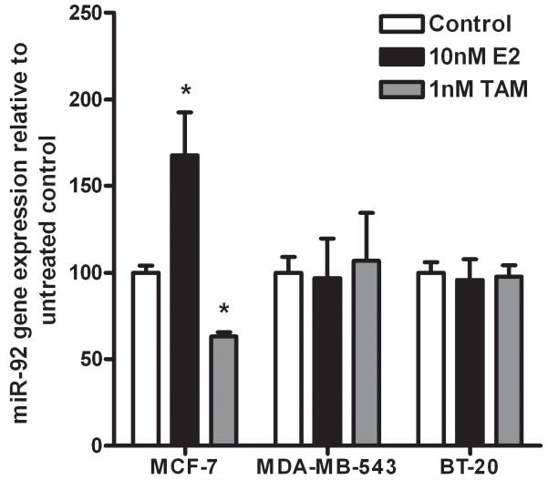
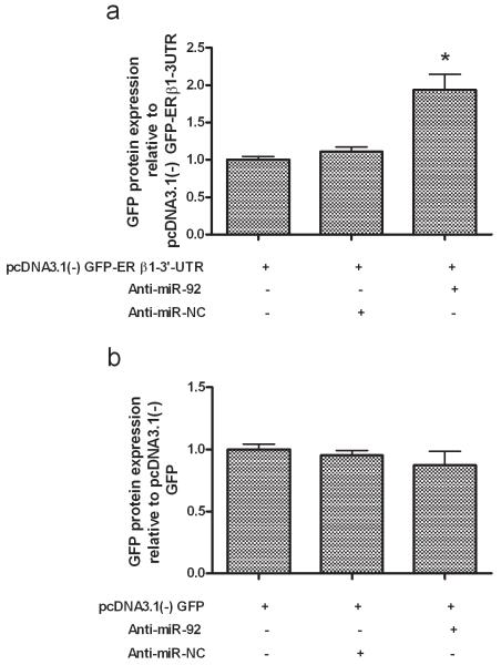
Estrogen receptor beta1 (ERbeta1) downregulation occurs in many breast cancers, but the responsible molecular mechanisms remain unclear. Here, we report that levels of ERbeta1 expression are negatively regulated by the microRNA miR-92. Expression analysis in a cohort of primary breast tumors confirmed a significant negative correlation between miR-92 and both ERbeta1 mRNA and protein. Inhibition of miR-92 in MCF-7 cells increased ERbeta1 expression in a dose-dependent manner, whereas miR-92 overexpression led to ERbeta1 downregulation. Reporter constructs containing candidate miR-92 binding sites in the 3'-untranslated region (UTR) of ERbeta1 suggested by bioinformatics analysis confirmed that miR-92 downregulated ERbeta1 via direct targeting of its 3'-UTR. Our results define a potentially important mechanism for downregulation of ERbeta1 expression in breast cancer.


Figures

Figure 1
Figure 1. Ratios of expression of ERβ1/miR-92 in breast cell lines and clinical breast samples
Quantitative RT-PCR analysis showed an inverse relationship between ERβ1 and miR-92in 4 breast cell lines of differing ER status (shown above each bar; a). A similar inverse correlation was observed in matched normal (N) and breast tumours (T), with high ratios in normal breast tissue and low ratios in breast tumours (b). In a subgroup of these samples this relationship was not observed with miR-124a (c), indicating specificity. Each experiment was performed in triplicate with 3 experimental replicates. Bars represent mean ± S.D.
Figure 2
Figure 2. miR-92 is negatively correlated with ERβ1 mRNA and protein in human breast cancer but not with ERα
Scatterplots showing an inverse correlation between expression of miR-92 and ERβ1 mRNA (a) determined by real-time RT-PCR (P = 0.001), and protein (b) determined by immunohistochemistry and Allred scoring (P = 0.04). Examples of ERβ1 immunohistochemistry and their relationship with miR-92 expression are shown in (c). No relationship was seen with ERα (d).
Figure 3
Figure 3. Effects of miR-92 manipulation on expression of ERβ1 and other target genes
Using quantitative RT-PCR, suppression of miR-92 inhibits miR-92 gene expression in MCF-7 cells in a dose-dependent manner and after 48 hours resulted in upregulation of ERβ1 mRNA expression relative to negative controls (a) while overexpression of miR-92 led to downregulation of ERβ1 mRNA expression (b). MiR-92 silencing restored MUC16 expression (c). Each experiment was performed in triplicate with 3 experimental replicates. Each data point is the mean ± S.D. *P < 0.05, **P <0.001, ***P < 0.0001.
Figure 4
Figure 4. Hormonal regulation of miR-92 expression
Quantitative RT-PCR analysis showed inhibition of miR-92 expression by TAM and its upregulation by E2 in MCF-7 cells but not in BT-20 and MDA-MB-453. Values are fold expression compared to vehicle control (EtOH) for miR-92. Each experiment was performed in triplicate with 3 experimental replicates. Each data point is the mean ± S.D. *P < 0.05.
Figure 5
Figure 5. ERβ1 is targeted by miR-92 via its 3′UTR in MCF-7 cells
MCF-7 cells were transiently transfected with plasmids to allow over-expression of GFP transcripts with either 3′-UTRs containing sequence from the ERβ1 3′-UTR including the potential miR-92 sites, or with unspecialised 3′-UTRs. Transfections also included either anti-miR-92 or a non-targeting control (NC). Reduction of endogenous miR-92 by anti-miR-92 led to an increase in GFP protein expression specified by the ERβ1 3′-UTR (a) and was not observed with the plasmid containing the unspecialised 3′-UTRs (b). Each experiment was performed in triplicate with 3 experimental replicates. Bars represent mean ± S.D. *P < 0.05.

References

    1. Fox EM, Davis RJ, Shupnik MA. ERβ in breast cancer-onlooker, passive player, or active protector? Steroids. 2008;73:1039–51. - PMC - PubMed
    1. Leung YK, Mak P, Hassan S, Ho SM. Estrogen receptor (ER)-β isoforms: a key to understanding ERβ signaling. Proc Natl Acad Sci U S A. 2006;103:13162–7. - PMC - PubMed
    1. Speirs V, Skliris GP, Burdall SE, et al. Distinct expression patterns of ERα and ERβ in normal human mammary gland. J Clin Pathol. 2002;55:371–4. - PMC - PubMed
    1. Treeck O, Lattrich C, Springwald A, et al. Estrogen receptor β exerts growth inhibitory effects on human mammary epithelial cells. Breast Cancer Res Treat. 2009 May 12; DOI 10.1007/s10549–009-0413–2. - PubMed
    1. Skliris GP, Munot K, Bell SM, et al. Reduced expression of oestrogen receptor beta in invasive breast cancer and its re-expression using DNA methyl transferase inhibitors in a cell line model. J Pathol. 2003;201:213–20. - PubMed

Publication types