. 2010 Jul 23;285(30):23420-32.
doi: 10.1074/jbc.M110.102830. Epub 2010 May 18.

Vinculin is a dually regulated actin filament barbed end-capping and side-binding protein

Affiliations

Vinculin is a dually regulated actin filament barbed end-capping and side-binding protein

Christophe Le Clainche et al. J Biol Chem. .

Abstract

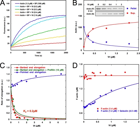
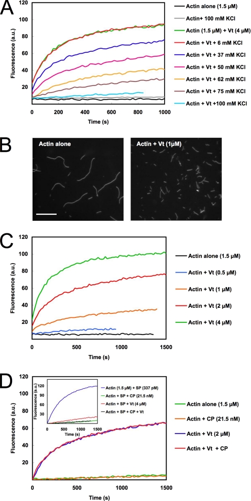
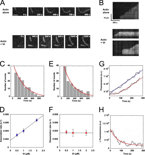
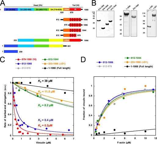
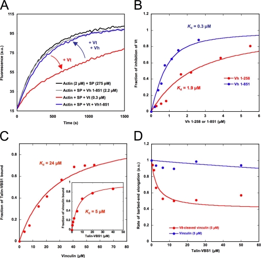
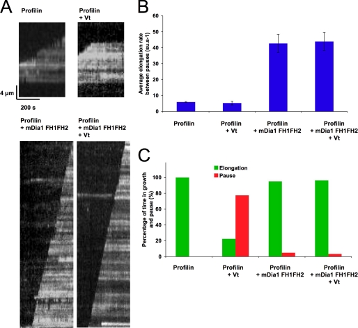
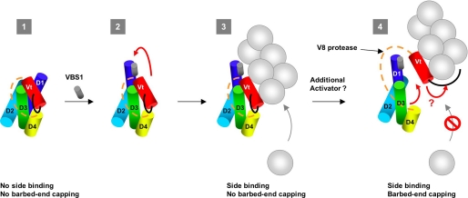
The focal adhesion protein vinculin is an actin-binding protein involved in the mechanical coupling between the actin cytoskeleton and the extracellular matrix. An autoinhibitory interaction between the N-terminal head (Vh) and the C-terminal tail (Vt) of vinculin masks an actin filament side-binding domain in Vt. The binding of several proteins to Vh disrupts this intramolecular interaction and exposes the actin filament side-binding domain. Here, by combining kinetic assays and microscopy observations, we show that Vt inhibits actin polymerization by blocking the barbed ends of actin filaments. In low salt conditions, Vt nucleates actin filaments capped at their barbed ends. We determined that the interaction between vinculin and the barbed end is characterized by slow association and dissociation rate constants. This barbed end capping activity requires C-terminal amino acids of Vt that are dispensable for actin filament side binding. Like the side-binding domain, the capping domain of vinculin is masked by an autoinhibitory interaction between Vh and Vt. In contrast to the side-binding domain, the capping domain is not unmasked by the binding of a talin domain to Vh and requires the dissociation of an additional autoinhibitory interaction. Finally, we show that vinculin and the formin mDia1, which is involved in the processive elongation of actin filaments in focal adhesions, compete for actin filament barbed ends.


Figures

FIGURE 1.
FIGURE 1.
The tail domain of vinculin inhibits actin filament barbed end elongation. A, barbed end growth was measured in the presence of 100 pm spectrin-actin seeds (SP), 1.5 μm MgATP-G-actin (10% pyrenyl-labeled), and the indicated concentrations of Vt. B, actin was polymerized in the presence of 100 pm spectrin-actin seeds, 1.5 μm MgATP-G-actin, and the indicated concentrations of Vt for 30 min. The polymerized (P) and unpolymerized (S) fractions were separated by ultracentrifugation, resolved by 10% SDS-PAGE, detected by Coomassie Blue staining (inset), and quantified. C, barbed end growth was measured in the presence of 100 pm spectrin-actin seeds, 1.5 μm MgATP-G-actin (10% pyrenyl-labeled), and increasing concentrations of Vt in the absence or presence of 12 μm profilin. Pointed end growth was measured in the presence of 4 nm gelsolin-actin complex, 2 μm MgATP-G-actin (10% pyrenyl-labeled), and increasing concentrations of Vt. The initial rate of pointed end growth and the rate of barbed end growth at 25% polymerization were plotted versus the concentration of Vt. D, Vt increases the concentration of F-actin at steady state in the presence of gelsolin but has no effect in the absence of gelsolin. 1.4 μm MgATP-F-actin (10% pyrenyl-labeled) was incubated overnight with increasing amounts of Vt in the absence or presence of gelsolin. All of the experiments were performed three times with the same results. a.u., arbitrary units.
FIGURE 2.
FIGURE 2.
Vinculin tail nucleates actin filaments that are capped at their barbed ends. A, spontaneous nucleation of 1.5 μm MgATP-G-actin (10% pyrenyl-labeled) was measured in the presence of 4 μm Vt, in a buffer containing 5 mm Tris, pH 7.8, 0.2 mm ATP, 1 mm MgCl2, 0.1 mm CaCl2, 1 mm DTT, and 10 mm KCl supplemented with the indicated concentrations of KCl. B, observation of single actin filaments nucleated by Vt in fluorescence microscopy. The polymerization of 5 μm MgATP-G-actin was measured in a buffer containing 5 mm Tris, pH 7.8, 0.2 mm ATP, 1 mm MgCl2, 0.1 mm CaCl2, 1 mm DTT, and 25 mm KCl in the absence (left) or presence of 1 μm Vt (right). As soon as 90% polymerization was reached, actin filaments were stabilized and stained with a molar equivalent of Alexa488-labeled phalloidin, diluted, and observed by fluorescence microscopy. Scale bar, 10 μm. C, the polymerization of 1.5 μm MgATP-G-actin (10% pyrenyl-labeled) was measured in the presence of increasing concentrations of Vt in a buffer containing 5 mm Tris, pH 7.8, 0.2 mm ATP, 1 mm MgCl2, 0.1 mm CaCl2, 1 mm DTT, and 25 mm KCl. D, Vinculin remains bound to the barbed end of nucleated filaments at low ionic strength. 1.5 μm MgATP-G-actin (10% pyrenyl-labeled) was polymerized alone, in the presence of 2 μm Vt or 21.5 nm CP or Vt and CP, in a buffer containing 5 mm Tris, pH 7.8, 0.2 mm ATP, 1 mm MgCl2, 0.1 mm CaCl2, 1 mm DTT, and 25 mm KCl. Inset, as a control, the effect of 21.5 nm CP or 4 μm Vt or Vt and CP on the elongation of 337 pm spectrin-actin seeds (SP) is shown in the same buffer conditions. All of the experiments were performed three times with the same results. a.u., arbitrary units.
FIGURE 3.
FIGURE 3.
Direct real-time observation of actin filament barbed end capping by vinculin tail. Conditions were as follows. 0.6 μm MgATP-G-actin (10% Alexa488-labeled) was polymerized in a flow chamber coated with NEM-myosin in 5 mm Tris, pH 7.8, 100 mm KCl, 0.2 mm ATP, 0.1 mm CaCl2, 1 mm MgCl2, 10 mm DTT, 0.2 mm 1,4-diazabicyclo(2,2,2)-octane, 0.2% methyl-cellulose. A, time lapse of the elongation of single actin filaments in the absence (top) or presence of 0.5 μm Vt (bottom). See also the corresponding supplemental Movies 1 and 2. Scale bar, 5 μm. B, kymographs of the single actin filaments shown in A. Actin filament fluorescence intensity was measured along its length (vertical axis) for each frame of the time lapse (horizontal axis). The filaments are oriented with the non-growing pointed end to the bottom and the growing barbed end to the top. In contrast with the continuous elongation observed for control filaments (top), barbed end growth was occasionally interrupted by pauses in the presence of Vt (bottom). C–F, quantification of the association and dissociation rate constants that characterized the interaction between Vt and actin filament barbed ends using TIRF microscopy. C and E, periods of growth and arrest were measured on kymographs corresponding to the elongation of actin filaments in the presence of 0.6 μm MgATP-G-actin (10% Alexa488-labeled) and 0.5 μm Vt. The exponential fit of the time distribution of the periods of growth (C) and arrest (E) gave the association and dissociation rates, respectively. The association rates (D) and dissociation rates (F) were plotted versus increasing concentrations of Vt. Error bars, S.E. G, measurement of the dissociation rate of vinculin from actin filament barbed ends after dilution by using a fluorescence bulk assay. A reaction in which spectrin-actin seeds have been pre-elongated in the presence of 2 μm actin and 2 μm Vt was diluted 20 times in a solution containing 2 μm MgATP-G-actin, 10% pyrenyl-labeled (red line). As a control, we performed the dilution of the same reaction, but we let Vt dissociate for 15 min before adding G-actin (blue line). H, the subtraction of the dissociation kinetics from the linear fit of the control kinetics shown in G gave the kinetics of dissociation of Vt from the barbed end. The exponential fit of the curve gave a dissociation rate constant k = 0.014 s−1. All of the experiments were performed three times with the same results. a.u., arbitrary units.
FIGURE 4.
FIGURE 4.
Identification of the critical domains of vinculin involved in the barbed end capping and F-actin side binding activities. A, constructs used in this study. B, SDS-PAGE of the purified proteins used in this work. C, barbed end growth was measured in the presence of 100 pm spectrin-actin seeds (SP), 1.5 μm MgATP-G-actin (10% pyrenyl-labeled), and the indicated concentrations of vinculin full-length (residues 1–1066), ΔD1 (residues 253–1066), 812–1066, 812–1044, 812–978, and 879–1066. The rate of barbed end growth at 25% polymerization was plotted versus the concentration of each vinculin construct. D, the co-sedimentation of vinculin 812–1066 (2 μm), 812–1044 (2 μm), 812–978 (2 μm), 253–1066 (2 μm), and full-length vinculin 1–1066 (2 μm) with F-actin was measured in the presence of increasing concentrations of F-actin. The fraction of vinculin bound to F-actin was plotted versus the concentration of actin. All of the experiments were performed three times with the same results. a.u., absorbance units.
FIGURE 5.
FIGURE 5.
Actin filament side binding and barbed end capping are both autoinhibited but regulated differently. A, vinculin head domain 1–851 (D1–D4) inhibits the capping activity of Vt. Barbed end growth was measured in the presence of 275 pm spectrin-actin seeds (SP), 2 μm MgATP-G-actin (10% pyrenyl-labeled), 0.3 μm Vt, and 2.2 μm vinculin head (residues 1–851). B, vinculin head domain 1–851 inhibits the capping activity of Vt more efficiently than Vh 1–258. Barbed end growth was measured in the presence of 100 pm spectrin-actin seeds, 2 μm MgATP-G-actin (10% pyrenyl-labeled), 0.75 μm Vt, and increasing amounts of vinculin head 1–258 and 1–851. The fraction of inhibition of Vt by Vh was plotted versus the concentration of each Vh construct. The best fit of the data gave the affinity of the head domain for the capping domain located in Vt. C, the VBS1 domain of talin binds to full-length vinculin with low affinity. The increase in fluorescence anisotropy of 3 μm IAEDANS-labeled VBS1 observed in the presence of increasing concentrations of full-length vinculin was taken as a measure of the VBS1-vinculin complex formation. The fraction of VBS1 bound to vinculin was plotted versus the total concentration of vinculin, and the best fit of the data gave a Kd of 24 μm. Inset, the affinity of full-length vinculin for VBS1 is enhanced by F-actin. The fraction of vinculin bound to VBS1 was obtained by measuring the fraction of vinculin (2 μm) that co-sedimented with F-actin (12 μm) in the presence of increasing concentrations of VBS1. The fraction of vinculin bound to VBS1 was plotted versus the concentration of VBS1, and the best fit of the data gave a Kd of 5 μm. D, the capping domain of vinculin, cleaved by V8 protease, is unmasked by the binding of the VBS1 domain of talin. Barbed end growth was measured in the presence of 100 pm spectrin-actin seeds, 2 μm MgATP-G-actin (10% pyrenyl-labeled), 5 μm intact full-length vinculin (blue points), or V8-cleaved vinculin (red points) and increasing concentrations of VBS1. All of the experiments were performed three times with the same results. a.u., absorbance units.
FIGURE 6.
FIGURE 6.
Vinculin tail and mDia1 compete for actin filament barbed ends. Conditions were as follows. 0.6 μm MgATP-G-actin (10% Alexa488-labeled) was polymerized in a flow chamber coated with NEM-myosin in 5 mm Tris pH 7.8, 100 mm KCl, 0.2 mm ATP, 0.1 mm CaCl2, 1 mm MgCl2, 10 mm DTT, 0.2 mm 1,4-diazabicyclo(2,2,2)-octane, 0.2% methyl-cellulose. A, representative kymographs of growing single actin filaments in the presence of 6 μm profilin (top left; see corresponding supplemental Movie 4); 6 μm profilin and 1 μm Vt (top right; see corresponding supplemental Movie 5); 20 nm mDia1-FH1FH2 and 6 μm profilin (bottom left; see corresponding supplemental Movie 6); or 20 nm mDia1-FH1FH2, 6 μm profilin, and 1 μm Vt (bottom right; see corresponding supplemental Movie 7). B, average elongation rates between the pauses in the indicated conditions. C, percentage of time in growth and pause in the indicated conditions. Error bars, S.E.
FIGURE 7.
FIGURE 7.
Working model for the regulation of vinculin. In this scheme, each subdomain of vinculin is represented as a cylinder, according to Refs. and . The proline-rich region that links Vh and Vt is represented as an orange dotted line. The C-terminal arm is represented as a black line (1). Vinculin exists in an autoinhibited conformation in which the F-actin binding site located in Vt is buried by contacts with the head subdomain D1. However, according to the crystal structure of vinculin and affinity measurements, Vt also interacts with the subdomains D3 and D4 (2). The low affinity binding of the VBS1 domain of talin is sufficient to disrupt the D1-Vt interaction (red arrow). However, other contacts between Vt and Vh remain (3). The exposed Vt domain binds to the side of actin filaments. In this conformation, vinculin allows barbed end elongation. Contacts between Vh and Vt still exist (4). In addition to the binding of VBS1 to D1, the disruption of unidentified contacts, probably between subdomains D3-D4 and Vt, leads to the full dissociation of Vt from Vh. This dissociation allows the C-terminal arm of Vt to cap the barbed end of the filament or change its structure to prevent the association of actin monomers.

References

    1. Ziegler W. H., Liddington R. C., Critchley D. R. (2006) Trends Cell Biol. 16, 453–460 - PubMed
    1. Zamir E., Geiger B. (2001) J. Cell Sci. 114, 3583–3590 - PubMed
    1. Coll J. L., Ben-Ze'ev A., Ezzell R. M., Rodríguez Fernández J. L., Baribault H., Oshima R. G., Adamson E. D. (1995) Proc. Natl. Acad. Sci. U.S.A. 92, 9161–9165 - PMC - PubMed
    1. Volberg T., Geiger B., Kam Z., Pankov R., Simcha I., Sabanay H., Coll J. L., Adamson E., Ben-Ze'ev A. (1995) J. Cell Sci. 108, 2253–2260 - PubMed
    1. Xu W., Coll J. L., Adamson E. D. (1998) J. Cell Sci. 111, 1535–1544 - PubMed

Publication types

MeSH terms