Comparative Study
. 2010 Aug 5;116(5):767-71.
doi: 10.1182/blood-2009-11-251926. Epub 2010 May 18.

Sezary syndrome and mycosis fungoides arise from distinct T-cell subsets: a biologic rationale for their distinct clinical behaviors

Affiliations
Comparative Study

Sezary syndrome and mycosis fungoides arise from distinct T-cell subsets: a biologic rationale for their distinct clinical behaviors

James J Campbell et al. Blood. .

Abstract

Cutaneous T-cell lymphoma (CTCL) encompasses leukemic variants (L-CTCL) such as Sézary syndrome (SS) and primarily cutaneous variants such as mycosis fungoides (MF). To clarify the relationship between these clinically disparate presentations, we studied the phenotype of T cells from L-CTCL and MF. Clonal malignant T cells from the blood of L-CTCL patients universally coexpressed the lymph node homing molecules CCR7 and L-selectin as well as the differentiation marker CD27, a phenotype consistent with central memory T cells. CCR4 was also universally expressed at high levels, and there was variable expression of other skin addressins (CCR6, CCR10, and CLA). In contrast, T cells isolated from MF skin lesions lacked CCR7/L-selectin and CD27 but strongly expressed CCR4 and CLA, a phenotype suggestive of skin resident effector memory T cells. Our results suggest that SS is a malignancy of central memory T cells and MF is a malignancy of skin resident effector memory T cells.


Figures
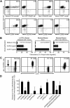
Figure 1
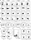
Figure 1
Malignant T cells from the blood of patients with L-CTCL have a central memory T-cell phenotype. (A) Flow cytometric analysis of clonal malignant T cells isolated from the blood of an L-CTCL patient with a TCR-Vβ17 malignant clone demonstrated that malignant T cells expressed high levels of the central memory T-cell markers L-selectin and CCR7. CCR4 was also expressed at high levels, but expression of other skin homing addressins (CLA, CCR6, and CCR10) was variable. There was no detectable expression of the gut homing addressins α4β7-integrin and CCR9. (B) CCR4 and CCR7 expressed by malignant T cells in L-CTCL were functional as demonstrated by their ability to support migration to the CCR4 ligand CCL22 and the CCR7 ligand CCL21 in in vitro T-cell migration assays. Shown are the mean ± SD migration of clonal malignant T cells from 3 L-CTCL donors (left panel) and the naive (center panel) and CLA+ memory T cells (right panel) from 3 normal donors. As expected, normal naive T cells responded only to CCR7 ligand. (C) Clonal malignant T cells from L-CTCL patients expressed high levels of CD27, consistent with a central memory T-cell phenotype, but expression of CD45RA varied among patients. CD4+ CD3+ T cells from a normal donor are shown on the left. The CD27-negative effector T-cell population observed in normal donors is indicated by an arrow. The right 3 panels show 3 L-CTCL patients, and histograms are gated to show only the CD4+ clonal malignant T-cell populations. CD27 was uniformly expressed on malignant cells from all donors, but the expression of CD45RA was variable. (D) Analysis of 11 additional L-CTCL patients with identifiable malignant clones produced similar results. Shown are the mean ± SD of surface marker expression of the CD3+/CD4+ cells expressing the expanded TCR-Vβ clonotype.
Figure 2
Figure 2
CD4+ T cells from MF skin lesions have an effector memory phenotype, but T cells from L-CTCL skin lesions have central memory characteristics. (A) Normal skin and the skin lesions of patients with L-CTCL contained a clear population of L-selectin and CCR7 coexpressing central memory T cells. However, T cells isolated from the skin lesions of stable patch plaque MF lacked coexpression of the central memory markers CCR7/L-selectin but did express the skin homing addressins CCR4 and CLA. All histograms are gated to show only CD4+ T cells. CCR4 was expressed by virtually all T cells, whereas CLA expression was frequent but not universal. Similar to results in L-CTCL patients, expression of CD45RO and CD45RA varied among patients. Four representative patients are shown; similar results were obtained in a total of 15 patients. (B) Clonal malignant T cells in the skin lesions of patients with L-CTCL expressed both L-selectin/CCR7 and skin homing addressins. Malignant T cells from the skin lesions of patients with L-CTCL were selectively studied by gating on T cells expressing the malignant Vβ subfamily (black cells, Vβ1 for patient 333 and Vβ13.1 for patient 330). Histograms are gated to show only malignant T cells. Malignant clonal T cells showed near-universal expression of the central memory markers L-selectin and CCR7 as well as high expression of the skin homing addressins CLA and CCR4. Results from 2 patients are shown; similar findings were observed in T cells isolated from the skin lesions of 4 additional L-CTCL patients. (C) In contrast, clonal T cells arising in MF skin lesions lacked central memory markers. Clonal malignant T cells expressing TCR-Vβ13.1 were evident in the skin lesions of a patient with MF (black cells represent remaining histograms gated to display only clonal malignant cells). Clonal malignant T cells lacked expression of the central memory markers L-selectin/CCR7 but expressed CCR4, and the majority coexpressed CLA. (D) Microarray gene expression analysis demonstrated that T cells from MF skin lesions expressed low levels of CD27 compared with clonal malignant T cells isolated from the blood of L-CTCL patients. The mean ± SEM of 5 MF patients, 3 normal skin patients, and 3 L-CTCL patients are shown. (E) Flow cytometric staining of T cells from the skin lesions of MF and L-CTCL confirmed that CD4+ T cells in MF show loss of CD27, consistent with an effector memory phenotype. T cells from the skin lesion of a representative patient with L-CTCL expressed CD27.

References

    1. Willemze R, Jaffe ES, Burg G, et al. WHO-EORTC classification for cutaneous lymphomas. Blood. 2005;105(10):3768–3785. - PubMed
    1. Shin J, Monti S, Aires DJ, et al. Lesional gene expression profiling in cutaneous T-cell lymphoma reveals natural clusters associated with disease outcome. Blood. 2007;110(8):3015–3027. - PMC - PubMed
    1. Kim YH, Liu HL, Mraz-Gernhard S, Varghese A, Hoppe RT. Long-term outcome of 525 patients with mycosis fungoides and Sezary syndrome: clinical prognostic factors and risk for disease progression. Arch Dermatol. 2003;139(7):857–866. - PubMed
    1. van Doorn R, van Kester MS, Dijkman R, et al. Oncogenomic analysis of mycosis fungoides reveals major differences with Sezary syndrome. Blood. 2009;113(1):127–136. - PubMed
    1. Clark RA, Chong BF, Mirchandani N, et al. A novel method for the isolation of skin resident T cells from normal and diseased human skin. J Invest Dermatol. 2006;126(5):1059–1070. - PubMed

Publication types

MeSH terms