. 2010 Aug 19;116(7):1165-71.
doi: 10.1182/blood-2009-12-255810. Epub 2010 May 18.

Th17/Treg ratio in human graft-versus-host disease

Affiliations

Th17/Treg ratio in human graft-versus-host disease

Philippe Ratajczak et al. Blood. .

Abstract

Th17 cells have never been explored in human graft-versus-host disease (GVHD). We studied the correlation between the presence of Th17 cells with histologic and clinical parameters. We first analyzed a cohort of 40 patients with GVHD of the gastrointestinal tract. Tumor necrosis factor (TNF), TNF receptors, and Fas expression, and apoptotic cells, CD4(+)IL-17(+) cells (Th17), and CD4(+)Foxp3(+) cells (Treg) were quantified. A Th17/Treg ratio less than 1 correlated both with the clinical diagnosis (P < .001) and more than 2 pathologic grades (P < .001). A Th17/Treg ratio less than 1 also correlated with the intensity of apoptosis of epithelial cells (P = .03), Fas expression in the cellular infiltrate (P = .003), TNF, and TNF receptor expression (P < .001). We then assessed Th17/Treg ratio in 2 other independent cohorts; a second cohort of 30 patients and confirmed that Th17/Treg ratio less than 1 correlated with the pathologic grade of GI GVHD. Finally, 15 patients with skin GVHD and 11 patients with skin rash but without pathologic GVHD were studied. Results in this third cohort of patients with skin disease confirmed those found in patients with GI GVHD. These analyses in 96 patients suggest that Th17/Treg ratio could be a sensitive and specific pathologic in situ biomarker of GVHD.


Figures

Figure 1
Figure 1. Th17 and Treg distribution in gut biopsies of patients with mild and severe GvHD
Th17 and Treg were identified by the expression of IL-17 or Foxp3, respectively in CD4+ cells using 2-colour immunohistochemistry (IL-17 in brown: A, B; Foxp3 in brown C, D; and CD4 in red; A,B,C,D) Th17 cells were more numerous in patients with mild GvHD (arrowhead: A) whereas Treg were present in patients with severe GvHD (arrowhead: D) Counts of double immunostained cells were independently assessed by 2 pathologists (PR and AJ) on an Olympus AX 70 microscope with wide-field eyepiece number 26.5. At 400× magnification, this wide-field eyepiece provided a field size of 0.344 mm2.
Figure 2
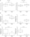
Figure 2. CD4+counts, CD4+Foxp3+ and CD4+Foxp3+ cell proportions in duodenal biopsies of patients with grade <2 or ≥ 2 (1st and 2nd cohorts)
A, Absolute number of CD4+ cells per field in the first and second cohorts of duodenal biopsie B, Percentage of CD4+ cells expressing Foxp3 in the same cohort C, Percentage of CD4+ cells expressing IL-17 in the same cohort An increased proportion of CD4+ cells express Foxp3 in duodenal biopsies of patients with grade ≥ 2 compared with patients with grade <2 in the two cohorts A decreased proportion of CD4+ cells express IL-17 in duodenal biopsies of patients with grade ≥ 2 compared with patients with grade <2 in the two cohorts.
Figure 3
Figure 3. CD8+ and CD8+Foxp3+ cells in duodenal biopsies in the 1st and 2nd cohorts
Number of CD8+ and CD8+Foxp3+ cells in GvHD patients with grade <2 or ≥ 2. While Foxp3 has a central role in Treg development, it is also clear that Foxp3 upregulation occurs with T cell activation. Thus, as a control of the specificity, CD8/Foxp3 double staining was performed on the two first cohorts. Counts showed that most of the CD8+ cells are not activated in the 2 independent cohorts of patients who underwent duodenal biopsies.
Figure 4
Figure 4. Double immunofluorescent staining with IL-17 and Foxp3 in skin biopsies of patients with acute, chronic and no GvHD
A, Skin biopsies showed infiltrates and apoptotic bodies (arrows) in acute and chronic lichenoid eruption but not in patients without GvHD. B, Overlay of double immunofluorescent staining with IL-17 (red) and Foxp3 (green) antibodies showed Foxp3 expressing cells in skin biopsies of patients with acute and chronic GVHD and IL17-expressing cells in skin biopsies of patients without GVHD. C, Enlargement of the stained cells showed that no of them expressed both IL- 17 and Foxp3.
Figure 5
Figure 5. Flow cytometry analyses of Th17, and of Tregs in the peripheral blood of 31 patients with acute, chronic or no chronic GvHD
Flow cytometry analyses of Th17, and of Tregs in the peripheral blood of 23 patients with acute, chronic or no chronic GvHD PBMCs from healthy controls (n = 5) and patients with GvHD (acute, n=12 and chronic, n=3) or without (n=8) were stained with anti-CD3, anti-CD4, anti-CD25 antibodies followed by intracellular Foxp3 antibody and examined by flow cytometry. Intracellular IL-17 antibody was also examined by flow cytometry after stimulation for 6 hours with PMA and Ionomycine. A, Frequencies of Tregs in patients and controls B, Frequencies of Th17 T cells in patients and controls C, Th17/Tregs ratio in patients and controls The mean value of each group is represented (—).

References

    1. Socie G, Blazar BR. Acute graft-versus-host disease; from the bench to the bedside. Blood. 2009 Nov 12;114(20):4327–4336. - PMC - PubMed
    1. Bettelli E, Korn T, Oukka M, Kuchroo VK. Induction and effector functions of T(H)17 cells. Nature. 2008 Jun 19;453(7198):1051–1057. - PMC - PubMed
    1. Bettelli E, Oukka M, Kuchroo VK. T(H)-17 cells in the circle of immunity and autoimmunity. Nat Immunol. 2007 Apr;8(4):345–350. - PubMed
    1. Kappel LW, Goldberg GL, King CG, et al. IL-17 contributes to CD4-mediated graft-versus-host disease. Blood. 2008 Oct 17; - PMC - PubMed
    1. Laurence A, O’Shea JJ. T(H)-17 differentiation: of mice and men. Nat Immunol. 2007 Sep;8(9):903–905. - PubMed

Publication types

MeSH terms