Review
. 2010 Oct 6;7 Suppl 5(Suppl 5):S631-47.
doi: 10.1098/rsif.2010.0115.focus. Epub 2010 May 19.

Biomimetic coatings for bone tissue engineering of critical-sized defects

Affiliations
Review

Biomimetic coatings for bone tissue engineering of critical-sized defects

Yuelian Liu et al. J R Soc Interface. .

Abstract

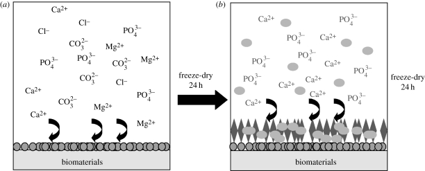
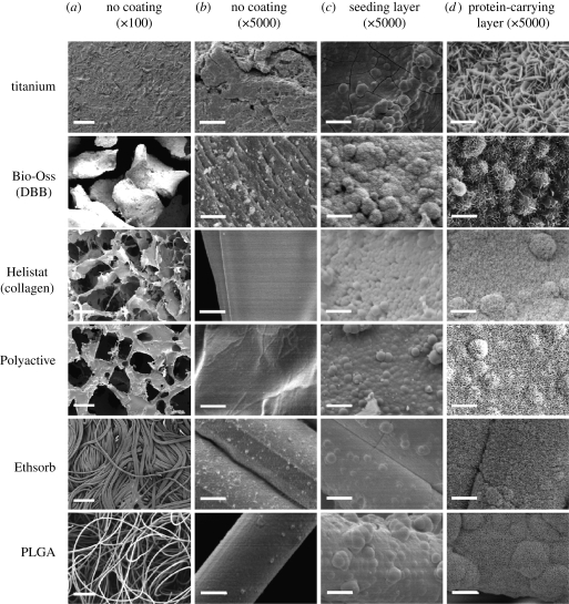
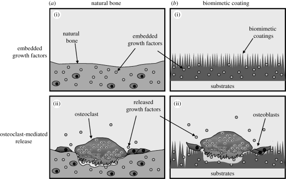
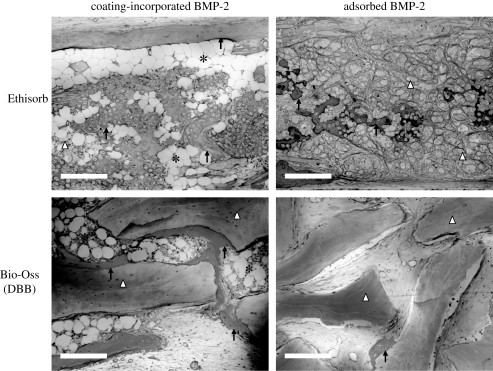
The repair of critical-sized bone defects is still challenging in the fields of implantology, maxillofacial surgery and orthopaedics. Current therapies such as autografts and allografts are associated with various limitations. Cytokine-based bone tissue engineering has been attracting increasing attention. Bone-inducing agents have been locally injected to stimulate the native bone-formation activity, but without much success. The reason is that these drugs must be delivered slowly and at a low concentration to be effective. This then mimics the natural method of cytokine release. For this purpose, a suitable vehicle was developed, the so-called biomimetic coating, which can be deposited on metal implants as well as on biomaterials. Materials that are currently used to fill bony defects cannot by themselves trigger bone formation. Therefore, biological functionalization of such materials by the biomimetic method resulted in a novel biomimetic coating onto different biomaterials. Bone morphogenetic protein 2 (BMP-2)-incorporated biomimetic coating can be a solution for a large bone defect repair in the fields of dental implantology, maxillofacial surgery and orthopaedics. Here, we review the performance of the biomimetic coating both in vitro and in vivo.
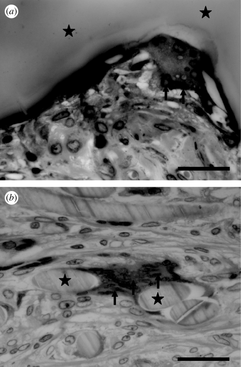

Figures

Figure 1.
Figure 1.
The procedure of the biphasic biomimetic calcium phosphate coating. (a) Step 1: the biomaterial is immersed in a fivefold concentrated simulated body fluid for 24 h at 37°C, 150 r.p.m. with stirring. A fine, dense layer of amorphous calcium phosphate (ACaP) thereby formed serves as a seeding substratum for the deposition of a more substantial crystalline layer. (b) Step 2: after freeze-drying, the crystalline layer will be produced by immersing the ACaP-coated biomaterials in a supersaturated calcium phosphate solution for 48 h at 37°C, 60 r.p.m. with shaking. The samples are then freeze-dried. If the crystalline layer is to be functionalized by the incorporation of bioactive agents such as bone morphogenetic protein-2 (BMP-2), then these agents are introduced into the latter medium at a certain concentration. Circles, amorphous seedling layer; ellipsoids, BMP-2; diamonds, crystalline protein-carrying layer.
Figure 2.
Figure 2.
(a) Scanning electron micrographs of the geometry at low magnifications and (b) the topography at high magnifications of six biomaterials in the native condition, and (c) after coating first with an amorphous seeding layer of calcium phosphate and (d) then with a crystalline one, which was coprecipitated with bovine serum albumin (BSA). The six materials include: one with metallic material titanium plates; one with bovine-derived mineral material Bio-Oss granules (deproteinized bovine bone, DBB); one with bovine-derived organic sponge Helistat (collagen); and three with synthetic polymeric membranes (Polyactive, Ethisorb and PLGA) (from top to bottom). Scale bars, (a) 200 µm and (bd) 5 µm. (Part of this figure was reproduced with kind permission from the Journal of Tissue Engineering and Mary Ann Liebert, Inc., New Rochelle, NY).
Figure 3.
Figure 3.
(a)(i,ii) Graph depicting both osteoclastic release of pre-embedded growth factors in bone matrix during bone remodelling and (b)(i,ii) the osteoclast-mediated release of growth factors that were incorporated into the calcium phosphate coatings of biomaterials.
Figure 4.
Figure 4.
Light micrographs of cross sections through the samples of Ethisorb (as a sample of a polymeric material) and Bio-Oss (deproteinized bovine bone, DBB) in the two groups (coated polymer bearing an incorporated depot of bone morphogenetic protein-2 (coating-incorporated BMP-2); uncoated polymer bearing an adsorbed depot of BMP-2 (adsorbed BMP-2)), five weeks after implantation in rats and stained with McNeil's tetrachrome, basic fuchsin and toluidine blue O. Scale bars, 200 µm. Newly regenerated bone tissue (indicated by black arrows) was extensively deposited along the surface of the two materials when they were functionalized by coating-incorporated BMP-2; a tremendous amount of bone marrow tissue (black asterisks) filled in the interstitial space. In contrast, bone tissue was sporadically deposited along the surface of the two materials that had an adsorbed depot of BMP-2; bone marrow tissue was rarely found in this group.
Figure 5.
Figure 5.
Light micrographs of cross sections through discs of the two polymer types ((a) polyactive; (b) PLGA in the group of unfunctionalized uncoated polymer only), five weeks after subcutaneous implantation in rats and stained with McNeil's tetrachrome, basic fuchsin and toluidine blue O. Scale bars, 30 µm. (a) The surface of Polyactive (indicated by black asterisks) was tightly covered by a spindle-like FBGC with several nuclei (indicated by black arrows) and ruffled edges. (b) Elliptic or round cross-sectioned PLGA fibres (indicated by black asterisks) sporadically distributed in connective tissues. In the middle of the image, a PLGA fibre was embraced and tightly covered by a FBGC with more than 10 nuclei (indicated by black arrows).

References

    1. Aggarwal S., Pittenger M. F. 2005. Human mesenchymal stem cells modulate allogeneic immune cell responses. Blood 105, 1815–1822. (10.1182/blood-2004-04-1559) - DOI - PubMed
    1. Allen T. D., Dexter T. M., Simmons P. J. 1990. Marrow biology and stem cells. Immunol. Ser. 49, 1–38. - PubMed
    1. Anderson J. M. 2000. Multinucleated giant cells. Curr. Opin. Hematol. 7, 40–47. (10.1097/00062752-200001000-00008) - DOI - PubMed
    1. Arm D. M., Tencer A. F., Bain S. D., Celino D. 1996. Effect of controlled release of platelet-derived growth factor from a porous hydroxyapatite implant on bone ingrowth. Biomaterials 17, 703–709. (10.1016/0142-9612(96)86740-8) - DOI - PubMed
    1. Asamura S., Ikada Y., Matsunaga K., Wada M., Isogai N. 2010. Treatment of orbital floor fracture using a periosteum–polymer complex. J. Craniomaxillofac. Surg. 38, 197–203. (10.1016/j.jcms.2009.06.011) - DOI - PubMed

MeSH terms

LinkOut - more resources