Review
. 2010 Sep;2(9):a004887.
doi: 10.1101/cshperspect.a004887. Epub 2010 May 19.

p63 and p73, the ancestors of p53

Affiliations
Review

p63 and p73, the ancestors of p53

V Dötsch et al. Cold Spring Harb Perspect Biol. 2010 Sep.

Abstract

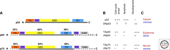
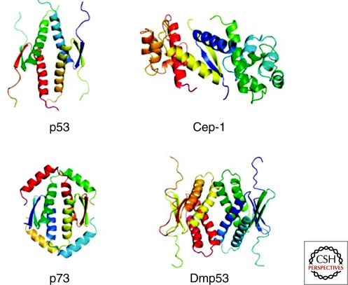
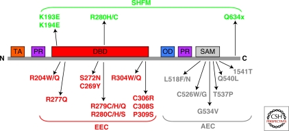
p73 and p63 are two homologs of the tumor suppressive transcription factor p53. Given the high degree of structural similarity shared by the p53 family members, p73 and p63 can bind and activate transcription from the majority of the p53-responsive promoters. Besides overlapping functions shared with p53 (i.e., induction of apoptosis in response to cellular stress), the existence of extensive structural variability within the family determines unique roles for p63 and p73. Their crucial and specific functions in controlling development and differentiation are well exemplified by the p63 and p73 knockout mouse phenotypes. Here, we describe the contribution of p63 and p73 to human pathology with emphasis on their roles in tumorigenesis and development.


Figures

Figure 1.
Figure 1.
Schematic representation of the protein modular structure of the p53 family members. (A) The p53 family includes three genes that encode p53, p73, and p63. The overall domain structure of p53, p73, and p63 is conserved and consists of an amino-terminal transactivation domain (TAD), a central DNA binding domain (DBD) and a carboxy-terminal oligomerization domain (OD). The longest α isoform of p73 and p63 contains a sterile alpha motif (SAM), a putative protein–protein interaction domain found in many signaling proteins and transcription factors. Identity shared by p73 and p63 with p53 is indicated. (B) Simplified classification of the functions of the p53 family members. Members of this family are involved in similar as well as in unique cellular functions, with the most important role of p63 and p73 being regulation of differentiation and development. Their crucial and specific functions in controlling development and differentiation are well exemplified by the p63 and p73 knockout mouse developmental phenotypes (C).
Figure 2.
Figure 2.
Structures of the ODs of p53, Cep-1 (C. elegans), p73 and Dmp53 (Drosophila melanogaster). The p53 OD is a tetramer of dimers in which each monomer contributes one β-strand and one α-helix. Dimers are assembled by the formation of an intermolecular antiparallel β-sheet, which is stabilized by hydrophobic interactions with two helices that also arrange in an antiparallel orientation. The tetramer is created by hydrophobic interactions between the helices of both dimers. In the case of p73 this arrangement is further stabilized by a second helix that reaches across the tetramerization interface. In the Drosophila protein Dmp53, every secondary structure element is doubled, and in Cep-1, tetramerization is inhibited by charged amino acids in the tetramerization interface. The dimeric structure of Cep-1 is stabilized by interaction with a SAM domain (one for each monomer).
Figure 3.
Figure 3.
Role of p63 in epidermal formation. (A) The epidermis is maintained throughout adult life by stem cells, which self-renew and produce progeny (TA cells) that undergoes terminal differentiation. TA and stem cells are located in the epidermal basal layer. A simplified scheme of the actions of p63 in epidermal development is shown here. (B) Several studies have been performed to identify the target promoters of p63. Here is shown a simplified and limited list of published p63 target genes that have been experimentally validated. The scheme does not necessarily imply that other target genes may be less important.
Figure 4.
Figure 4.
Regulation of developmental sympathetic neuronal survival and apoptosis by the p53 family members. The p53 protein family has been implicated in the death signaling following NGF withdrawal. ΔNp73 is an NGF-induced anti-apoptotic protein in sympathetic neurons. (A) During development, neuronal survival is guaranteed by the availability of NGF, which in turn maintains the ΔNp73 levels high enough to inhibit the pro-apoptotic functions of both p53 and TAp63. (B) On the other hand, once the levels of NGF becomes restrictive, neuronal cells are committed to cell death through down-regulation of ΔNp73 and augmented TAp63. Under these circumstances, the antagonistic action of ΔNp73 is released, and the proapototic family members can elicit cell death by initiating mitochondrial apoptosis.
Figure 5.
Figure 5.
p63 mutations in human syndromes. p63 mutations in the EEC and AEC syndromes cluster respectively in DBD and SAM domains. Conserved protein domains are shown for TAp63: orange, TA domain; red, DNA-binding domain (DBD); blu, isomerization domain (OD); purple, proline-rich regions (PR); gray, SAM domain. Heterozygous missense mutations identified in EEC, AEC, and SHFM syndromes are indicated. All AEC mutations are located in the SAM domain, whereas all missense mutations in EEC syndrome are in the DNA-binding domain.

References

    1. Adorno M, Cordenonsi M, Montagner M, Dupont S, Wong C, Hann B, Solari A, Bobisse S, Rondina MB, Guzzardo V, et al.2009. A Mutant-p53/Smad complex opposes p63 to empower TGFβ-induced metastasis. Cell 137: 87–98 - PubMed
    1. Andreas CJ, Sridharan R, Eviatar N, Dmitry BV, Carol VR, et al.2009. Structural evolution of p53, p63, and p73: Implication for heterotetramer formation. PNAS, in press - PMC - PubMed
    1. Barbieri CE, Tang LJ, Brown KA, Pietenpol JA 2006. Loss of p63 leads to increased cell migration and up-regulation of genes involved in invasion and metastasis. Cancer Res 66: 7589–7597 - PubMed
    1. Belyi VA, Levine AJ 2009. One billion years of p53/p63/p73 evolution. Proc Natl Acad Sci 106: 17609–17610 - PMC - PubMed
    1. Billon N, Terrinoni A, Jolicoeur C, McCarthy A, Richardson WD, Melino G, Raff M 2004. Roles for p53 and p73 during oligodendrocyte development. Development 131: 1211–1220 - PubMed