

Review
doi: 10.1101/cshperspect.a000588.
Epub 2010 May 19.
Gene positioning
Affiliations
- PMID: 20484389
- PMCID: PMC2869523
- DOI: 10.1101/cshperspect.a000588
Item in Clipboard
Review
Gene positioning
Cold Spring Harb Perspect Biol.
2010 Jun.
Abstract
Eukaryotic gene expression is an intricate multistep process, regulated within the cell nucleus through the activation or repression of RNA synthesis, processing, cytoplasmic export, and translation into protein. The major regulators of gene expression are chromatin remodeling and transcription machineries that are locally recruited to genes. However, enzymatic activities that act on genes are not ubiquitously distributed throughout the nucleoplasm, but limited to specific and spatially defined foci that promote preferred higher-order chromatin arrangements. The positioning of genes within the nuclear landscape relative to specific functional landmarks plays an important role in gene regulation and disease.
Figures
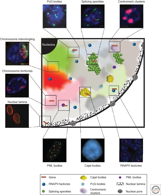
Nuclear subcompartments. An illustration of the mammalian cell nucleus showing nuclear domains and features of gene positioning (orange bar) that have been identified thus far. The nuclear positioning of genes and their associations with different nuclear landmarks are implicated in gene activation and gene repression as discussed in the text. DNA counterstain in blue where applicable. Centromeric clusters. Human CD2 transgenes (green) colocalize with a centromeric cluster (red) in nonexpressing T cells from hCD2 transgenic mice (line 1.3A14). Reprinted by permission from (Hiragami-Hamada et al. 2009). Splicing speckles. The C3/C4 genomic region of murine chromosome 8 (red) associates with splicing speckles (green) in fetal liver cells. Reprinted by permission from (Noordermeer et al. 2008). PcG bodies. Bxd gene (red) positioned at PcG bodies (green) in the posterior part of wild-type stage 5-8 hour Drosophila embryos examined by 2D-FISH. Reprinted by permission from Macmillan Publishers Ltd: Nature Cell Biology (Lanzuolo et al. 2007), copyright (2007). Chromosome intermingling. Intermingling between chromosomes 5 (green) and 7 (red) in human lymphocytes. Reproduced with permission from (Branco and Pombo 2006). Chromosome territories. uPA locus (green) is positioned inside of its chromosome territory (CT; chromosome 10, red) in HepG2 cells. Reproduced with permission from (Ferrai et al. 2010). Nuclear lamina. Igh locus (green) interaction with the nuclear lamina (LMNB1, red) in NIH3T3. Reprinted by permission from Macmillan Publishers Ltd: Nature (Reddy et al. 2008), copyright (2008). PML bodies. MHCII region (red) is proximal to PML bodies (green) in control fibroblasts. Reproduced with permission from (Shiels et al. 2001). Cajal bodies. Simultaneous hybridization of RNU1, RNU2, RNU3, RNU7, RNU12, HIST1, and HIST2 loci show several simultaneous gene interactions with Cajal bodies (red). Reprinted from (Frey et al. 1999). Copyright (1999), with permission from Elsevier. RNAPII factories. Association of hCD2 transgenes with RNAPII-S2p in T cells from hCD2 transgenic mouse line 1.3B (SQ Xie, R Festenstein, A Pombo, unpubl.).
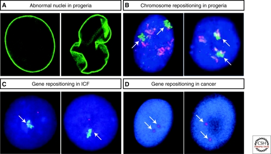
Nuclear organization defects in disease. (A) Abnormal nuclear shape in Hutchinson-Gilford Progeria Syndrome. Nuclei in patients with progeria show an abnormal morphology (right) contrasting with the regular nuclei shape found in healthy individuals (left). Reproduced with permission from (Scaffidi et al. 2005). (B) Chromosome repositioning in Hutchinson-Gilford Progeria Syndrome. Interior repositioning of human chromosome 13 (green, arrows) in a HGPS fibroblast cell line (right), in comparison to a control cell line (left). Proliferation marker pKi-67 (red). Reproduced with permission from (Meaburn et al. 2007). (C) Gene repositioning in immunodeficiency centromeric instability facial anomalies (ICF). In control male cells (left), the SYBL1 (red, arrows) inactive allele (Y chromosome) is localized inside or at the edge of the Y CT, whereas in ICF cells (right) it loops out of its CT and escapes silencing. Reproduced with permission from (Matarazzo et al. 2007). Copyright (2007) National Academy of Sciences. (D) Gene repositioning in early mammary tumorigenesis. Position of the cancer AKT1 locus (red, arrows) before (left) and after (right) oncogenic activation of an epithelial cell line. After activation there is a radial shift in the locus position toward the periphery. Reproduced with permission from (Meaburn and Misteli 2008).
References
-
- Akhtar A, Gasser SM 2007. The nuclear envelope and transcriptional control. Nat Rev Genet 8:507–517 - PubMed
-
- Amano T, Sagai T, Tanabe H, Mizushina Y, Nakazawa H, Shiroishi T 2009. Chromosomal dynamics at the Shh locus: Limb bud-specific differential regulation of competence and active transcription. Dev Cell 16:47–57 - PubMed
-
- Andrulis ED, Neiman AM, Zappulla DC, Sternglanz R 1998. Perinuclear localization of chromatin facilitates transcriptional silencing. Nature 394:592–595 - PubMed
Publication types
MeSH terms
Substances
Grants and funding
LinkOut - more resources
Full Text Sources