. 2010 Jul;24(7):1453-68.
doi: 10.1210/me.2010-0087. Epub 2010 May 19.

cAMP-dependent activation of mammalian target of rapamycin (mTOR) in thyroid cells. Implication in mitogenesis and activation of CDK4

Affiliations

cAMP-dependent activation of mammalian target of rapamycin (mTOR) in thyroid cells. Implication in mitogenesis and activation of CDK4

Sara Blancquaert et al. Mol Endocrinol. 2010 Jul.

Abstract

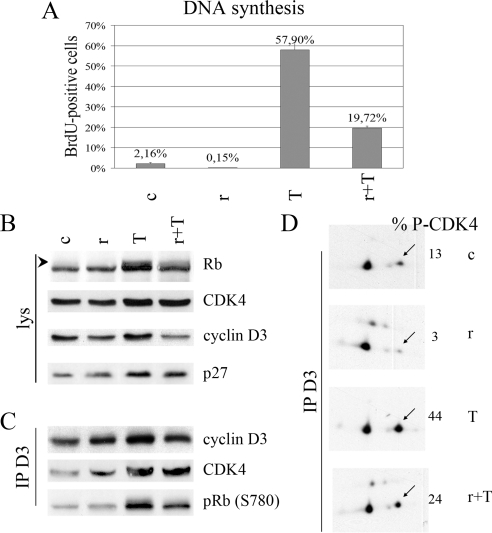
How cAMP-dependent protein kinases [protein kinase A (PKA)] transduce the mitogenic stimulus elicited by TSH in thyroid cells to late activation of cyclin D3-cyclin-dependent kinase 4 (CDK4) remains enigmatic. Here we show in PC Cl3 rat thyroid cells that TSH/cAMP, like insulin, activates the mammalian target of rapamycin (mTOR)-raptor complex (mTORC1) leading to phosphorylation of S6K1 and 4E-BP1. mTORC1-dependent S6K1 phosphorylation in response to both insulin and cAMP required amino acids, whereas inhibition of AMP-activated protein kinase and glycogen synthase kinase 3 enhanced insulin but not cAMP effects. Unlike insulin, TSH/cAMP did not activate protein kinase B or induce tuberous sclerosis complex 2 phosphorylation at T1462 and Y1571. However, like insulin, TSH/cAMP produced a stable increase in mTORC1 kinase activity that was associated with augmented 4E-BP1 binding to raptor. This could be caused in part by T246 phosphorylation of PRAS40, which was found as an in vitro substrate of PKA. Both in PC Cl3 cells and primary dog thyrocytes, rapamycin inhibited DNA synthesis and retinoblastoma protein phosphorylation induced by TSH and insulin. Although rapamycin reduced cyclin D3 accumulation, the abundance of cyclin D3-CDK4 complexes was not affected. However, rapamycin inhibited the activity of these complexes by decreasing the TSH and insulin-mediated stimulation of activating T172 phosphorylation of CDK4. We propose that mTORC1 activation by TSH, at least in part through PKA-dependent phosphorylation of PRAS40, crucially contributes to mediate cAMP-dependent mitogenesis by regulating CDK4 T172-phosphorylation.


Figures

Figure 1
Figure 1
TSH and forskolin induce p70S6K1 phosphorylation but do not activate PKB. PC Cl3 cells were treated without (c) or with 5 μg/ml insulin (i) and/or 10 μm forskolin (F) or 1 mU/ml TSH (T) for 20 min. In some conditions (in panel B) cells were pretreated with 200 nm wortmannin (w) before stimulation. PKB activity was assayed after immunoprecipitation by incubation with a specific substrate and [γ32P] ATP. 32P incorporation in PKB substrate was measured by scintillation counting (CPM). In parallel, immunoblotting after SDS-PAGE from the same samples was performed for detection of PKB, pPKB (S473), pPKB (T308), S6K1 and pS6K1 (T389). A and B, Two independent experiments are illustrated.
Figure 2
Figure 2
TSH and insulin induce phosphorylation of S6K1 and 4E-BP1. A, Kinetics of S6K1 phosphorylation stimulated by insulin and/or TSH. PC Cl3 cells were treated or not (−) with 5 μg/ml insulin (i) and/or 1 mU/ml TSH (T) for 5 min up to 20 h. After SDS-PAGE total S6K1 was detected by immunoblotting. Arrows and arrowheads indication the position of hypophosphorylated and hyperphosphorylated S6K1 forms, respectively. A (lower panel) shows the densitometry scanning quantitation of the T389 phosphorylation of S6K1 detected by immunoblotting from the same samples. Data are expressed as fold stimulation compared with untreated control cells. B–D, Rapamycin inhibits most but not all phosphorylation of S6K1 (B and C) and 4E-BP1 (D) induced by insulin and/or TSH. PC Cl3 cells were treated without (c) or with 5 μg/ml insulin (i) and/or 1 mU/ml TSH (T) for 20 min. Rapamycin, 40 nm (rapa or r) was added or not 2 h before treatment. B, S6K1, pS6K1 (T421/S424), pS6K1 (T389), pS6K1 (T229), and pS6 (S235/236) were immunodetected after SDS-PAGE of whole-cell lysates. C, S6K1 was immunoprecipitated (IP S6K1), the immunoprecipitate was separated by two-dimensional gel electrophoresis, and total S6K1 was detected by immunoblotting. The main nonmodified form of S6K1 is numbered as 0, and the presumably (multi-) phosphorylated forms are numbered from 1–13 according to their increasing isoelectric point shifts. D, 4E-BP1, p4E-BP1 (T37/S46), p4E-BP1 (S65), and pmTOR (S2448) were immunodetected after SDS-PAGE of whole-cell lysates.
Figure 3
Figure 3
TSH and cAMP increase the phosphorylation of PRAS40 but not of TSC2. PC Cl3 cells were treated without (c) or with 5 μg/ml insulin (i) and/or 10 μm forskolin (F) or 1 mU/ml TSH (T) for 20 min. pS6K1 (T389), p4E-BP1 (T37/46), pTSC2 (T1462), pTSC2 (Y1571), TSC2 and pPRAS40 (T246), were immunodetected after SDS-PAGE of whole-cell lysates.
Figure 4
Figure 4
cAMP agonists as well as insulin activate mTORC1 by stimulating the binding of 4E-BP1 to raptor and the phosphorylation of PRAS40. In panels A–C, PC Cl3 cells were treated without (−) or with (+) 10 μm forskolin (F) for 20 min. In panels D and E, they were treated without (c) or with 5 μg/ml insulin (i) and/or 10 μm forskolin (F) or 1 mU/ml TSH (T) for 20 min. A, Forskolin activates mTORC1. mTOR, raptor, and pS6K1 (T389) were immunodetected from whole-cell lysates (lys) (upper left panel). Endogenous mTOR complexes were immunoprecipitated (IP) with a raptor antibody (CRap) and a sample was taken for immunoblotting detection of mTOR and raptor (upper right panel). The immune complexes were mixed with [γ-32P]ATP and recombinant 4E-BP1 fragment, and the in vitro kinase reaction (K) was performed without (c) or with mTOR inhibitors, 10 μm LY294002 (LY), 5 μm FKBP12 (FKB), or FKBP12 + 5 μm rapamycin (FKB + r), and phosphorylation of 4E-BP1 was visualized by immunoblotting of p4E-BP1 (T37/46) or phosphorimaging (32P incorporation)(lower panel). B, Forskolin increases the T246 phosphorylation of PRAS40 and reduces the association of PRAS40 with mTORC1. mTOR, raptor, pS6K1 (T389), and pPRAS40 (T246) were immunodetected from whole-cell lysates (lys) (left panel). mTOR complexes were immunoprecipitated with the raptor antibody (CRap), and coimmunoprecipitated mTOR and PRAS40 were immunodetected (right panel). C, High-salt washing of mTORC1 increases its basal activity. mTOR complexes were immunoprecipitated with the raptor antibody (CRap) and washed with the same buffer containing 500 mm NaCl or 150 mm NaCl. Coimmunoprecipitated mTOR and raptor were immunodetected and the kinase activity (K) of these complexes was assayed as in panel A. D, Insulin, forskolin, and TSH activate mTORC1. mTORC1 complexes were immunoprecipitated with the raptor antibody (CRap) or nonimmune IgG (NI) as a control, and a sample was taken for immunoblotting detection of coimmunoprecipitated mTOR, raptor, and LST8. The kinase activity (K) of these complexes was assayed as in panel A. E, Insulin, forskolin, and TSH stimulate the binding of raptor to immobilized 4E-BP1. The same cell extracts as in panel D were incubated with 4E-BP1 or its mutant F113A (F/A) coupled to agarose beads. 4E-BP1-bound raptor was immunoblotted.
Figure 5
Figure 5
PRAS40 can be phosphorylated by PKA. A, Recombinant PRAS40 wild type (wt) and mutant T246A proteins (0.5 μg) were incubated with PKA catalytic subunit (0.1 μg), which was pretreated without or with H-89 (10 μm) or PKA inhibitor fragment 14–22 (10 μm) (PKI). Phosphorylation of PRAS40 was visualized by phosphoimager (32P incorporation) and T246 phosphorylation [pPRAS40(T246)] was immunodetected with a phosphospecific antibody. Substrates were stained with 0.5% ponceau S (PRAS40). 32P-labeled PRAS40 wt and T246A were digested by trypsin and resolved by two-dimensional phosphopeptide mapping with electrophoresis at pH 1.9 as the first dimension and chromatography as the second dimension. The peptide containing the T246 phosphorylation is shown (T246). B, PKA activation stimulates the phosphorylation of PRAS40, S6K1, and S6 in PC Cl3 cells and dog thyrocytes in primary culture. The cells were treated without (c) or with 5 μg/ml insulin (i) or the selective PKA activator N6-monobutyryl-cAMP (MB; 500 μm) for 20 min. pS6K1 (T389), S6K1, pS6 (S235/236), pPRAS40 (T246), and PRAS40 were immunodetected after SDS-PAGE of whole-cell lysates.
Figure 6
Figure 6
mTORC1 activity is involved in cell cycle progression, Rb phosphorylation, and cyclin D3-CDK4 activation stimulated by both insulin and TSH in PC Cl3 cells. Cells were stimulated without (c) or with 5 μg/ml insulin (i) and/or 1 mU/ml TSH (T) for 48 h (panel A) or 20 h (panels B–D) in the presence or not of 40 nm rapamycin (r). A, Rapamycin inhibits insulin- and TSH-stimulated DNA synthesis. BrdU was present during the last 24 h. The percentage of BrdU-positive nuclei was counted at the microscope. Results are mean + sd from four independent experiments. B, Rapamycin inhibits insulin- and TSH-stimulated Rb phosphorylation and cyclin D3 accumulation. Rb, pRb (T826), CDK4, cyclin D3, and p27 were immunodetected from whole-cell lysates (lys). The arrowhead indicates phosphorylated forms of Rb. C, Rapamycin inhibits insulin- and TSH-stimulated cyclin D3-CDK4 activity. Cell lysates were immunoprecipitated (IP) with anticyclin D3 (D3) or anti-p27 (p27) antibodies, assayed for Rb-kinase activity, separated by SDS-PAGE, and immunoblotted. Cyclin D3, CDK4, p27, and the in vitro T826 phosphorylation of the Rb fragment [pRb (T826)] were detected using specific antibodies. D, Rapamycin inhibits insulin- and TSH-stimulated activating phosphorylation of CDK4. Cyclin D3-CDK4 complexes were immunoprecipitated (IP) with cyclin D3 antibodies (D3). The immune complexes were separated by two-dimensional gel electrophoresis, and CDK4 was immunodetected. The percentage of the T172-phosphorylated form of CDK4 (arrows) vs. total CDK4 is indicated (% P-CDK4).
Figure 7
Figure 7
mTORC1 activity is involved in cAMP-dependent cell cycle progression and CDK4 activation in dog thyrocytes in primary culture. Dog thyrocytes cultured with 1 μg/ml insulin were stimulated without (c) or with 1 mU/ml TSH (T) for 48 h (panel A) or 20 h (panels B–D) in the presence or not of 40 nm rapamycin (r). A, Rapamycin inhibits TSH-induced DNA synthesis. BrdU was present during the last 24 h. The percentage of BrdU-positive nuclei was counted at the microscope (mean + range from duplicate dishes). B, Rapamycin inhibits TSH-stimulated Rb phosphorylation. Rb, CDK4, cyclin D3, and p27 were immunodetected from whole-cell lysates (lys). The arrowhead indicates phosphorylated forms of Rb. C, Rapamycin inhibits TSH-stimulated cyclin D3-CDK4 activity. Cell lysates were immunoprecipitated (IP) with anticyclin D3 (D3) antibodies, assayed for Rb-kinase activity, separated by SDS-PAGE, and immunoblotted. Cyclin D3, CDK4, and the in vitro S780 phosphorylation of the Rb fragment [pRb (S780)] were detected using specific antibodies. D, Rapamycin inhibits basal and TSH-stimulated activating phosphorylation of CDK4. Cyclin D3-CDK4 complexes were immunoprecipitated (IP) with cyclin D3 antibodies (D3). The immune complexes were separated by 2-dimensional gel electrophoresis, and CDK4 was immunodetected. The percentage of the T172-phosphorylated form of CDK4 (arrows) vs. total CDK4 is indicated (% P-CDK4).

Similar articles

Cited by

References

    1. Dumont JE, Maenhaut C, Christophe D, Vassart G, Roger PP 2005 Thyroid regulatory factors. In: DeGroot LJ, Jameson JL, eds. Endocrinology. New York: Elsevier Saunders; 1837–1860
    1. Roger PP, Servais P, Dumont JE 1983 Stimulation by thyrotropin and cyclic AMP of the proliferation of quiescent canine thyroid cells cultured in a defined medium containing insulin. FEBS Lett 157:323–329 - PubMed
    1. Roger P, Taton M, Van Sande J, Dumont JE 1988 Mitogenic effects of thyrotropin and adenosine 3′,5′-monophosphate in differentiated normal human thyroid cells in vitro. J Clin Endocrinol Metab 66:1158–1165 - PubMed
    1. Kimura T, Van Keymeulen A, Golstein J, Fusco A, Dumont JE, Roger PP 2001 Regulation of thyroid cell proliferation by thyrotropin and other factors : a critical evaluation of in vitro models. Endocr Rev 22:631–656 - PubMed
    1. Furuya F, Lu C, Willingham MC, Cheng SY 2007 Inhibition of phosphatidylinositol 3-kinase delays tumor progression and blocks metastatic spread in a mouse model of thyroid cancer. Carcinogenesis 28:2451–2458 - PubMed

Publication types

MeSH terms