. 2010 Aug;84(15):7703-12.
doi: 10.1128/JVI.02560-09. Epub 2010 May 19.

Upregulation of the chemokine (C-C motif) ligand 2 via a severe acute respiratory syndrome coronavirus spike-ACE2 signaling pathway

Affiliations

Upregulation of the chemokine (C-C motif) ligand 2 via a severe acute respiratory syndrome coronavirus spike-ACE2 signaling pathway

I-Yin Chen et al. J Virol. 2010 Aug.

Abstract

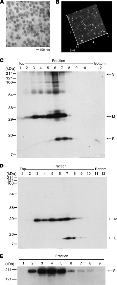
Severe acute respiratory syndrome coronavirus (SARS-CoV) was identified to be the causative agent of SARS with atypical pneumonia. Angiotensin-converting enzyme 2 (ACE2) is the major receptor for SARS-CoV. It is not clear whether ACE2 conveys signals from the cell surface to the nucleus and regulates expression of cellular genes upon SARS-CoV infection. To understand the pathogenesis of SARS-CoV, human type II pneumocyte (A549) cells were incubated with the viral spike protein or with SARS-CoV virus-like particles containing the viral spike protein to examine cytokine modulation in lung cells. Results from oligonucleotide-based microarray, real-time PCR, and enzyme-linked immunosorbent assays indicated an upregulation of the fibrosis-associated chemokine (C-C motif) ligand 2 (CCL2) by the viral spike protein and the virus-like particles. The upregulation of CCL2 by SARS-CoV spike protein was mainly mediated by extracellular signal-regulated kinase 1 and 2 (ERK1/2) and AP-1 but not the IkappaBalpha-NF-kappaB signaling pathway. In addition, Ras and Raf upstream of the ERK1/2 signaling pathway were involved in the upregulation of CCL2. Furthermore, ACE2 receptor was activated by casein kinase II-mediated phosphorylation in cells pretreated with the virus-like particles containing spike protein. These results indicate that SARS-CoV spike protein triggers ACE2 signaling and activates fibrosis-associated CCL2 expression through the Ras-ERK-AP-1 pathway.


Figures

FIG. 1.
FIG. 1.
Purification of SARS-CoV VLPs and the viral spike protein. The production of SARS-CoV VLPs in Sf9 cells was analyzed by electron microscopy (A) and AFM (B; the height image in liquid is shown). Western blot analysis was performed for identification of the SARS-CoV VLPs (C) and SARS-CoV(EM) VLPs (D) following purification by sucrose gradient centrifugation and for identification of the viral spike protein following Ni2+ Sepharose purification (E). Antibodies against the His and V5 tags of the recombinant viral proteins were used in Western blot analysis. Fractions 6 in panel C, 7 in panel D, and 4 in panel E were used in this study.
FIG. 2.
FIG. 2.
The elevation of CCL2 expression in SARS-CoV VLP-treated and SARS-CoV spike protein-treated A549 and Vero E6 cells. RNA was isolated from A549 cells at 24 h (A) or at indicated time points (B) after the treatment of purified SARS-CoV VLPs or SARS-CoV(EM) VLPs at a VLP-to-cell ratio of 10:1 or of the viral spike protein (1 μg/106 cells) as indicated. The level of CCL2 mRNA was determined by quantitative real-time PCR. In panel A, the CCL2 mRNA in Vero E6 cells was analyzed as a comparison. In addition, culture medium of the A549 cells was collected at 24 h after the treatment and subjected to ELISA to determine the level of secreted CCL2 (C). Cells without any of the treatments were analyzed in parallel as the controls. The results shown represent the means plus standard deviations (error bars) from four independent experiments. ***, P < 0.001; **, P < 0.01 (for comparisons between the treated and untreated control cells).
FIG. 3.
FIG. 3.
The involvement of AP-1 in the SARS-CoV VLP-induced upregulation of CCL2. (A) Illustration of the luciferase reporter plasmids containing the proximal regulatory region of the human CCL2 gene. pGLM-PRM-mT1T2 and pGLM-PRM-mκB plasmids that contain mutated (m) AP-1 and NF-κB binding sites are indicated. (B) Luciferase reporter assay. A549 cells were incubated for 24 h with SARS-CoV VLPs at 2 days posttransfection of the luciferase reporter plasmids, as indicated. Cell lysates were harvested and subjected to a luciferase reporter assay. The results shown represent the means plus standard deviations (error bars) from four independent experiments.
FIG. 4.
FIG. 4.
The increased expression of c-Fos and c-Jun in SARS-CoV VLP-treated A549 cells. Cell lysates prepared from A549 cells at various time points after treatment of SARS-CoV VLPs were subjected to Western blot analysis with antibodies against c-Fos, c-Jun, and tubulin as indicated. Samples indicated as 0 min represent controls in which cells were washed with fresh medium right after treatment with the SARS-CoV VLPs. Relative intensity (RI) shown was calculated by normalization of the intensities from the controls.
FIG. 5.
FIG. 5.
The role of MAPK pathway in the SARS-CoV-induced upregulation of CCL2 in A549 cells. A549 cells were treated with SARS-CoV VLPs (A) or with the viral spike protein (B) for various time periods. Cell lysates were then prepared and subjected to Western blot analysis with antibodies against phospho-ERK1/2 (p-ERK), ERK1/2 (ERK), phospho-JNK (p-JNK), JNK, phospho-p38 (p-p38), and p38 as indicated. Samples indicated as 0 min represent controls in which cells were washed with fresh medium right after treatment with the SARS-CoV VLPs or the viral spike protein as indicated. Relative phosphorylated levels of ERK, JNK, and p38 are shown.
FIG. 6.
FIG. 6.
The role of the MAPK pathway in SARS-CoV-induced upregulation of CCL2 in Vero E6 cells. Vero E6 cells were treated with SARS-CoV VLPs for various time periods. Cell lysates were then prepared and subjected to Western blot analysis with antibodies against phospho-ERK1/2 (p-ERK) and ERK1/2 (ERK) (A), phospho-JNK (p-JNK) and JNK (B), and phospho-p38 (p-p38) and p38 (C) as indicated. Samples marked time zero represent controls as described in the legend of Fig. 5.
FIG. 7.
FIG. 7.
The effects of ERK and JNK inhibitors on SARS-CoV-induced activation of MAPK. A549 cells were pretreated with MEK inhibitor PD98059 (20 μM; PD) or JNK inhibitor SP600125 (30 μM; SP) for 1 h before incubation with SARS-CoV VLPs (A) or with the viral spike protein (B) for 2 h. Cell lysates were prepared and subjected to Western blot analysis with antibodies against phospho-ERK1/2 (p-ERK), ERK1/2 (ERK), phospho-JNK (p-JNK), and JNK as indicated.
FIG. 8.
FIG. 8.
IκB-NF-κB signaling pathway is not involved in SARS-CoV-induced CCL2 expression. A549 cells were treated with SARS-CoV VLPs (A) or with the viral spike protein (B) for various time periods as indicated. Cell lysates were prepared and subjected to Western blot analysis with specific antibodies against IκBα and tubulin as indicated. Samples marked time zero represent controls as described in the legend to Fig. 5.
FIG. 9.
FIG. 9.
ERK1/2 regulate the promoter activity of CCL2 induced by SARS-CoV VLPs. At 2 days posttransfection of A549 cells with pGLM-PRM plasmid, the cells were treated with the inhibitor PD98059 (PD) or SP600125 (SP) for 1 h prior to a further incubation with SARS-CoV VLPs for 24 h. A luciferase reporter assay was performed. The results shown represent the means plus standard deviations (error bars) from four independent experiments. DMSO, dimethyl sulfoxide.
FIG. 10.
FIG. 10.
The effects of Ras and Raf inhibitors on the SARS-CoV-induced activation of ERK. A549 cells were pretreated with the Ras inhibitor manumycin A (MA; 3 μM), or Raf inhibitor GW5074 (GW; 1 μM) for 30 min prior to incubation with SARS-CoV VLPs (A) or with the viral spike protein (B) for 2 h. Cell lysates were prepared and subjected to Western blot analysis with antibodies against phospho-ERK1/2 (p-ERK) and ERK1/2 (ERK) as indicated. In addition, total RNA was isolated from A549 cells that had been treated with MAPK cascade inhibitors: MA and GW for 30 min and PD, SP, and p38 inhibitor SB203580 (SB) for 1 h, prior to the 6-h incubation with SARS-CoV VLPs or the viral spike protein. The level of CCL2 mRNA was determined by quantitative real-time PCR (C). The results shown represent the means plus standard deviations (error bars) from three independent experiments.
FIG. 11.
FIG. 11.
ACE2 is involved in the SARS-CoV-induced activation of ERK and upregulation of CCL2. (A) SARS-CoV spike protein upregulates the expression of CCL2 via the ACE2 signaling pathway. A549 cells were pretreated with anti-ACE2 antibody (Ab) or control IgG for 20 min prior to the incubation with SARS-CoV VLPs or the viral spike protein for 24 h. Total RNA was isolated for determination of the level of CCL2 mRNA by quantitative real-time PCR. The results shown represent the means plus standard deviations (error bars) from three independent experiments. (B) SARS-CoV spike protein activates ERK via the ACE2 signaling pathway. A549 cells were pretreated with anti-ACE2 antibody for 20 min prior to incubation with SARS-CoV VLPs or the viral spike protein for 2 h. Cell lysates were prepared and subjected to Western blot analysis with antibodies against phospho-ERK1/2 (p-ERK) and ERK1/2 (ERK) as indicated.
FIG. 12.
FIG. 12.
Phosphorylation of ACE2 in SARS-CoV VLP-treated Vero E6 cells. (A) The domain structure of ACE2. ACE2 is a type I integral membrane protein with a signal peptide (amino acid residues 1 to 17), an ectodomain (amino acid residues 18 to 740), a transmembrane domain (amino acid residues 741 to 763), and a cytoplasmic domain (amino acid residues 764 to 805). Potential phosphorylation sites for tyrosine kinase (TK; amino acid residues 775 to 781) and casein kinase II (CK II; amino acid residues 787 to 790) in the cytoplasmic domain are indicated. (B) Phosphorylated form of ACE2 in the SARS-CoV-permissive Vero E6 cells. Vero E6 cells were pretreated with the casein kinase II inhibitor DRB (100 μM) for 1 h prior to incubation with SARS-CoV VLPs for various times, as indicated. Cell lysates were prepared and subjected to immunoprecipitation (IP) with anti-phospho-Ser/Thr antibodies, followed by Western blot (WB) analysis with anti-ACE2 antibodies. Cell lysates without the process of immunoprecipitation (10% of input) were analyzed in parallel as loading controls. Lane C represents protein lysate prepared from untreated Vero E6 cells as an ACE2 positive control.
FIG. 13.
FIG. 13.
Critical role of casein kinase II in the SARS-CoV-induced ERK activation and upregulation of CCL2 in A549 cells. A549 cells were pretreated with the casein kinase II inhibitor DRB for 1 h. Following a 6-h incubation of the DRB-treated cells with SARS-CoV VLPs or the viral spike protein, total RNA was isolated to determine the level of CCL2 mRNA by quantitative real-time PCR (A). The results shown represent the means plus standard deviations (error bars) from four independent experiments. Meanwhile, cell lysates were prepared from the DRB-treated cells following a 2-h incubation with SARS-CoV VLPs or the viral spike protein and subjected to Western blot analysis with antibodies against phospho-ERK1/2 (p-ERK) and ERK1/2 (ERK) as indicated (B).
FIG. 14.
FIG. 14.
A current model of the ACE2 signaling pathway involved in SARS-CoV-induced CCL2 expression. Infection of lung epithelial cells with SARS-CoV induces casein kinase II (CK II)-mediated phosphorylation of the ACE2 receptor, leading to the activation of ERK1/2 and AP-1 and the upregulation of CCL2. Ras, Raf, and MEK may contribute to the SARS-CoV-induced ERK1/2 activation and CCL2 upregulation. The elevated level of CCL2 protein detected in the sera of SARS-CoV-infected patients may account for the development of lung fibrosis.

Similar articles

Cited by

References

    1. Castilletti, C., L. Bordi, E. Lalle, G. Rozera, F. Poccia, C. Agrati, I. Abbate, and M. R. Capobianchi. 2005. Coordinate induction of IFN-alpha and -gamma by SARS-CoV also in the absence of virus replication. Virology 341:163-169. - PMC - PubMed
    1. Chang, L., and M. Karin. 2001. Mammalian MAP kinase signalling cascades. Nature 410:37-40. - PubMed
    1. Chang, M. F., C. Y. Sun, C. J. Chen, and S. C. Chang. 1993. Functional motifs of delta antigen essential for RNA binding and replication of hepatitis delta virus. J. Virol. 67:2529-2536. - PMC - PubMed
    1. Chang, Y. J., C. Y. Liu, B. L. Chiang, Y. C. Chao, and C. C. Chen. 2004. Induction of IL-8 release in lung cells via activator protein-1 by recombinant baculovirus displaying severe acute respiratory syndrome-coronavirus spike proteins: identification of two functional regions. J. Immunol. 173:7602-7614. - PubMed
    1. Cheung, C. Y., L. L. Poon, I. H. Ng, W. Luk, S. F. Sia, M. H. Wu, K. H. Chan, K. Y. Yuen, S. Gordon, Y. Guan, and J. S. Peiris. 2005. Cytokine responses in severe acute respiratory syndrome coronavirus-infected macrophages in vitro: possible relevance to pathogenesis. J. Virol. 79:7819-7826. - PMC - PubMed

Publication types

MeSH terms