. 2010 Aug;84(15):7869-79.
doi: 10.1128/JVI.00485-10. Epub 2010 May 19.

The ubiquitin-proteasome system plays an important role during various stages of the coronavirus infection cycle

Affiliations

The ubiquitin-proteasome system plays an important role during various stages of the coronavirus infection cycle

Matthijs Raaben et al. J Virol. 2010 Aug.

Abstract

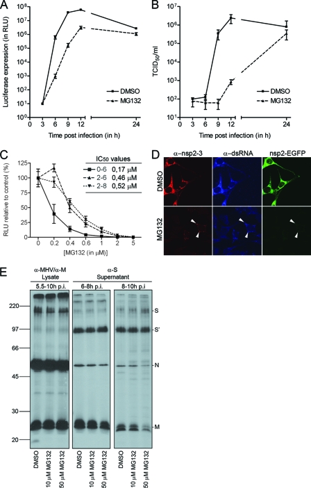
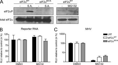
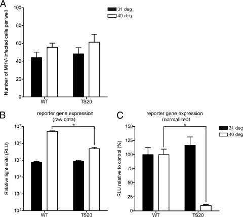
The ubiquitin-proteasome system (UPS) is a key player in regulating the intracellular sorting and degradation of proteins. In this study we investigated the role of the UPS in different steps of the coronavirus (CoV) infection cycle. Inhibition of the proteasome by different chemical compounds (i.e., MG132, epoxomicin, and Velcade) appeared to not only impair entry but also RNA synthesis and subsequent protein expression of different CoVs (i.e., mouse hepatitis virus [MHV], feline infectious peritonitis virus, and severe acute respiratory syndrome CoV). MHV assembly and release were, however, not appreciably affected by these compounds. The inhibitory effect on CoV protein expression did not appear to result from a general inhibition of translation due to induction of a cellular stress response by the inhibitors. Stress-induced phosphorylation of eukaryotic translation initiation factor 2alpha (eIF2alpha) generally results in impaired initiation of protein synthesis, but the sensitivity of MHV infection to proteasome inhibitors was unchanged in cells lacking a phosphorylatable eIF2alpha. MHV infection was affected not only by inhibition of the proteasome but also by interfering with protein ubiquitination. Viral protein expression was reduced in cells expressing a temperature-sensitive ubiquitin-activating enzyme E1 at the restrictive temperature, as well as in cells in which ubiquitin was depleted by using small interfering RNAs. Under these conditions, the susceptibility of the cells to virus infection was, however, not affected, excluding an important role of ubiquitination in virus entry. Our observations reveal an important role of the UPS in multiple steps of the CoV infection cycle and identify the UPS as a potential drug target to modulate the impact of CoV infection.


Figures

FIG. 1.
FIG. 1.
MHV infection is reduced by treatment with MG132. (A) LR7 cells were inoculated with MHV-EFLM (multiplicity of infection of 1) in the absence (DMSO control) or presence of 10 μM MG132. After 2 h, the inoculum was replaced with fresh medium containing either DMSO or 10 μM MG132. The luciferase expression levels at the indicated time points were determined. Standard deviations (n = 3) are indicated. (B) In parallel, viral infectivity in the culture media was determined by a quantal assay on LR7 cells. The 50% tissue culture infectious dose (TCID50) values are indicated. (C) LR7 cells were infected with MHV-EFLM in the absence (DMSO control) or presence of different concentrations of MG132. The drug was present from 0 to 6, from 2 to 6, or from 2 to 8 h postinfection. The intracellular luciferase expression levels were determined at the end of the treatment. Luciferase expression is indicated as a percentage relative to the DMSO control. The 50% inhibitory concentrations (IC50) for each condition are indicated in the graph. (D) MHV-nsp2EGFP-infected LR7 cells were mock treated or treated with 10 μM MG132 from 2 to 7 h postinfection. At 7 h postinfection, the cells were fixed and processed for immunofluorescence. The white arrowheads indicate the putative replication sites. (E) MHV-infected cells were labeled with 35S-labeled amino acids and subsequently chased. Progeny virions released into the culture medium from 6 to 8 and from 8 to 10 h postinfection (p.i.) were affinity purified using antibodies against the S protein. At 10 h postinfection, the cells were lysed and processed for immunoprecipitation with a mixture of anti-MHV and anti-M protein serum. Immunoprecipitates were analyzed by SDS-PAGE. Cells were mock treated or treated with 10 or 50 μM MG132 from 5.5 h postinfection onward. Molecular mass markers are indicated at the left in kilodaltons, while the positions of the MHV structural proteins (i.e., M, N, and S) in the gel are depicted at the right (S′ refers to the furin-cleaved forms of the S proteins).
FIG. 2.
FIG. 2.
The inhibitory effect of MG132 on MHV replication does not result from induction of a cellular stress response. (A) The phosphorylation status of eIF2α in the eIF2WT and eIF2S51A was determined by Western blotting with eIF2αP-specific antibodies (top) and related to total eIF2α levels by stripping and reprobing of the membrane (bottom). As a control, the cells were treated with 0.5 mM sodium arsenite (S.A.) for 30 min, which is known to induce the phosphorylation of eIF2α at serine 51 (36). In addition, the phosphorylation status of eIF2α in the eIF2WT MEFs was determined after treatment with 10 μM MG132 for 4 h. (B) The indicated cells (i.e., LR7, eIF2WT, and eIF2S51A) were transfected with a reporter RNA and subsequently cultured in the absence or presence of 10 μM MG132 (from 2 to 6 h posttransfection). The luciferase levels at 6 h posttransfection are indicated as a percentage relative to the DMSO control. (C) The different cells were infected with MHV-EFLM and subsequently cultured in the absence or presence of 10 μM MG132 (from 2 to 6 h postinfection). The intracellular luciferase expression levels at 6 h postinfection are indicated as a percentage relative to the DMSO control of each individual cell line. The standard deviations (n = 6) are indicated.
FIG. 3.
FIG. 3.
MHV infection is reduced by treatment with different proteasome inhibitors. (A) LR7 cells were infected with MHV-EFLM in the absence or presence of different concentrations of the inhibitors MG132, epoxomicin, lactacystin, and Velcade. The compounds were added either from 0 to 6 or from 2 to 6 h after virus inoculation (p.i.). The intracellular luciferase expression levels at 6 h postinfection are indicated as a percentage relative to the DMSO control. (B) LR7 cells transfected with the pEGFP-degron plasmid were treated with the indicated concentrations of the inhibitors for 6 h. Subsequently, the cells were fixed and processed for microscopic analysis. Representative images for each condition are shown. (C) LR7 cells infected with MHV-EFLM, FCWF cells infected with FIPV-Δ3abcFL, and Vero cells infected with SARS-CoV-GFP were treated with the indicated concentrations of Velcade. Velcade was applied to the cells 1 h prior to infection, after which the intracellular luciferase activity was measured at 7 h postinfection for MHV-EFLM and FIPV-Δ3abcFL. Quantification of GFP expression in Vero cells infected with SARS-CoV-GFP (53) was performed at 18 h postinfection by using a molecular light imager (Berthold Technologies). The reporter gene expression levels (i.e., FL for MHV and FIPV; GFP for SARS-CoV) are indicated as a percentage relative to the PBS control. The standard deviations are indicated (i.e., n = 3 for MHV and FIPV and n = 4 for SARS-CoV).
FIG. 4.
FIG. 4.
Velcade does not inhibit SARS-CoV main protease activity. A fluorimetric SARS-CoV nsp5 protease activity assay (see Materials and Methods) was performed. (A) Change in fluorescence as a result of cleavage of a Dabcyl-VRLQSGTC-fluorescein peptide substrate by wild-type nsp5 or an active site mutant (nsp5C145A) in the presence or absence of 50 μM Velcade. Symbols: □, wild-type nsp5; ○, wild-type nsp5 + Velcade; ▾, nsp5C145A. (B) Cleavage rate for wild-type nsp5 in the absence or presence of different concentrations of Velcade.
FIG. 5.
FIG. 5.
Replication but not entry of MHV is affected in cells deficient for ubiquitin conjugation. E36 (indicated as wild-type [WT]) or ts20 (indicated as TS20) Chinese hamster cells grown in 24-wells clusters were infected with MHV-EFLM at the indicated temperatures (i.e., 31 and 40°C). Incubation was continued at the same temperatures for 8 h. (A) Indicated are the total numbers of virus-infected cells per well for each condition, which were determined by staining for viral antigen with a polyclonal anti-MHV serum. (B and C) In parallel, the intracellular luciferase expression levels at 8 h postinfection were determined. The raw RLU values are shown in panel B, whereas in panel C the data are expressed as a percentage relative to the expression levels at 31 and 40°C in the wild-type cells (i.e., normalized data). The standard deviations are indicated (n = 6). *, P < 0.05 as determined by statistical analysis using the Student t test.
FIG. 6.
FIG. 6.
MHV replication is reduced in cells depleted of ubiquitin. HeLa-CEACAM1a cells grown in 96-wells clusters were transfected with 10 nM siRNAs targeting the indicated ubiquitin genes (i.e., UBA52 and Rps27A). Mock- and scrambled siRNA-transfected cells were used as controls. (A) At 72 h after transfection, the cells were lysed and processed for Western blotting with antibodies against ubiquitin and β-actin (loading control). The levels of free ubiquitin and ubiquitin conjugates were quantified and are expressed as a percentages relative to mock-transfected cells. Note that the values were corrected for the loading control. (B) The siRNA-transfected cells were inoculated with MHV-EFLM. At 6 h postinfection, the cells were fixed and stained for viral antigen using the polyclonal anti-MHV serum. The total numbers of MHV-infected cells per well are indicated, as well as the standard deviations (n = 3). (C) In parallel, the intracellular luciferase expression levels measured at 6 h postinfection are shown as a percentage relative to the control (scrambled siRNA-transfected cells). Note that the RLU values are corrected for cell viability. *, P < 0.05 as determined by statistical analysis using the Student t test.

Similar articles

Cited by

References

    1. Anand, K., J. Ziebuhr, P. Wadhwani, J. R. Mesters, and R. Hilgenfeld. 2003. Coronavirus main proteinase (3CLpro) structure: basis for design of anti-SARS drugs. Science 300:1763-1767. - PubMed
    1. Barretto, N., D. Jukneliene, K. Ratia, Z. Chen, A. D. Mesecar, and S. C. Baker. 2005. The papain-like protease of severe acute respiratory syndrome coronavirus has deubiquitinating activity. J. Virol. 79:15189-15198. - PMC - PubMed
    1. Berlanga, J. J., J. Santoyo, and C. De Haro. 1999. Characterization of a mammalian homolog of the GCN2 eukaryotic initiation factor 2α kinase. Eur. J. Biochem. 265:754-762. - PubMed
    1. Berlanga, J. J., I. Ventoso, H. P. Harding, J. Deng, D. Ron, N. Sonenberg, L. Carrasco, and C. de Haro. 2006. Antiviral effect of the mammalian translation initiation factor 2α kinase GCN2 against RNA viruses. EMBO J. 25:1730-1740. - PMC - PubMed
    1. Bieniasz, P. D. 2006. Late budding domains and host proteins in enveloped virus release. Virology 344:55-63. - PubMed

Publication types

MeSH terms