Redirecting lentiviral vectors pseudotyped with Sindbis virus-derived envelope proteins to DC-SIGN by modification of N-linked glycans of envelope proteins
- PMID: 20484510
- PMCID: PMC2898243
- DOI: 10.1128/JVI.00435-10
Redirecting lentiviral vectors pseudotyped with Sindbis virus-derived envelope proteins to DC-SIGN by modification of N-linked glycans of envelope proteins
Abstract
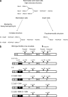
Redirecting the tropism of viral vectors enables specific transduction of selected cells by direct administration of vectors. We previously developed targeting lentiviral vectors by pseudotyping with modified Sindbis virus envelope proteins. These modified Sindbis virus envelope proteins have mutations in their original receptor-binding regions to eliminate their natural tropisms, and they are conjugated with targeting proteins, including antibodies and peptides, to confer their tropisms on target cells. We investigated whether our targeting vectors interact with DC-SIGN, which traps many types of viruses and gene therapy vectors by binding to the N-glycans of their envelope proteins. We found that these vectors do not interact with DC-SIGN. When these vectors were produced in the presence of deoxymannojirimycin, which alters the structures of N-glycans from complex to high mannose, these vectors used DC-SIGN as their receptor. Genetic analysis demonstrated that the N-glycans at E2 amino acid (aa) 196 and E1 aa 139 mediate binding to DC-SIGN, which supports the results of a previous report of cryoelectron microscopy analysis. In addition, we investigated whether modification of the N-glycan structures could activate serum complement activity, possibly by the lectin pathway of complement activation. DC-SIGN-targeted transduction occurs in the presence of human serum complement, demonstrating that high-mannose structure N-glycans of the envelope proteins do not activate human serum complement. These results indicate that the strategy of redirecting viral vectors according to alterations of their N-glycan structures would enable the vectors to target specific cells types expressing particular types of lectins.
Figures


Similar articles
-
Pseudotyping Lentiviral Vectors: When the Clothes Make the Virus.Viruses. 2020 Nov 16;12(11):1311. doi: 10.3390/v12111311. Viruses. 2020. PMID: 33207797 Free PMC article. Review.
-
Pseudotyping lentiviral vectors with aura virus envelope glycoproteins for DC-SIGN-mediated transduction of dendritic cells.Hum Gene Ther. 2011 Oct;22(10):1281-91. doi: 10.1089/hum.2010.196. Epub 2011 Jun 13. Hum Gene Ther. 2011. PMID: 21452926 Free PMC article.
-
Redirecting lentiviral vectors by insertion of integrin-tageting peptides into envelope proteins.J Gene Med. 2009 Jul;11(7):549-58. doi: 10.1002/jgm.1339. J Gene Med. 2009. PMID: 19434609 Free PMC article.
-
Differential N-linked glycosylation of human immunodeficiency virus and Ebola virus envelope glycoproteins modulates interactions with DC-SIGN and DC-SIGNR.J Virol. 2003 Jan;77(2):1337-46. doi: 10.1128/jvi.77.2.1337-1346.2003. J Virol. 2003. PMID: 12502850 Free PMC article.
-
Receptors and tropisms of envelope viruses.Curr Opin Virol. 2011 Jul;1(1):13-8. doi: 10.1016/j.coviro.2011.05.001. Curr Opin Virol. 2011. PMID: 21804908 Free PMC article. Review.
Cited by
-
Pseudotyping Lentiviral Vectors: When the Clothes Make the Virus.Viruses. 2020 Nov 16;12(11):1311. doi: 10.3390/v12111311. Viruses. 2020. PMID: 33207797 Free PMC article. Review.
-
Shaping viral immunotherapy towards cancer-targeted immunological cell death.Front Oncol. 2025 Jul 8;15:1540397. doi: 10.3389/fonc.2025.1540397. eCollection 2025. Front Oncol. 2025. PMID: 40697381 Free PMC article. Review.
-
Measles virus glycoprotein-pseudotyped lentiviral vectors are highly superior to vesicular stomatitis virus G pseudotypes for genetic modification of monocyte-derived dendritic cells.J Virol. 2012 May;86(9):5192-203. doi: 10.1128/JVI.06283-11. Epub 2012 Feb 15. J Virol. 2012. PMID: 22345444 Free PMC article.
-
Challenges in lentiviral vector production: retro-transduction of producer cell lines.Front Bioeng Biotechnol. 2025 May 29;13:1569298. doi: 10.3389/fbioe.2025.1569298. eCollection 2025. Front Bioeng Biotechnol. 2025. PMID: 40511300 Free PMC article.
-
Dendritic cell-specific intercellular adhesion molecule-3-grabbing nonintegrin (DC-SIGN) is a cellular receptor for delta inulin adjuvant.Immunol Cell Biol. 2024 Aug;102(7):593-604. doi: 10.1111/imcb.12774. Epub 2024 May 17. Immunol Cell Biol. 2024. PMID: 38757764 Free PMC article.
References
-
- Barry, S. C., B. Harder, M. Brzezinski, L. Y. Flint, J. Seppen, and W. R. Osborne. 2001. Lentivirus vectors encoding both central polypurine tract and posttranscriptional regulatory element provide enhanced transduction and transgene expression. Hum. Gene Ther. 12:1103-1108. - PubMed
-
- Betenbaugh, M. J., N. Tomiya, S. Narang, J. T. Hsu, and Y. C. Lee. 2004. Biosynthesis of human-type N-glycans in heterologous systems. Curr. Opin. Struct. Biol. 14:601-606. - PubMed
-
- Brown, B. D., A. Cantore, A. Annoni, L. S. Sergi, A. Lombardo, P. Della Valle, A. D'Angelo, and L. Naldini. 2007. A microRNA-regulated lentiviral vector mediates stable correction of hemophilia B mice. Blood 110:4144-4152. - PubMed
Publication types
MeSH terms
Substances
Grants and funding
LinkOut - more resources
Full Text Sources
Other Literature Sources

