. 2010 Sep;91(Pt 9):2203-15.
doi: 10.1099/vir.0.020958-0. Epub 2010 May 19.

Mutational analysis of the latency-associated nuclear antigen DNA-binding domain of Kaposi's sarcoma-associated herpesvirus reveals structural conservation among gammaherpesvirus origin-binding proteins

Affiliations

Mutational analysis of the latency-associated nuclear antigen DNA-binding domain of Kaposi's sarcoma-associated herpesvirus reveals structural conservation among gammaherpesvirus origin-binding proteins

Soo-Jin Han et al. J Gen Virol. 2010 Sep.

Abstract

The latency-associated nuclear antigen (LANA) of Kaposi's sarcoma-associated herpesvirus functions as an origin-binding protein (OBP) and transcriptional regulator. LANA binds the terminal repeats via the C-terminal DNA-binding domain (DBD) to support latent DNA replication. To date, the structure of LANA has not been solved. Sequence alignments among OBPs of gammaherpesviruses have revealed that the C terminus of LANA is structurally related to EBNA1, the OBP of Epstein-Barr virus. Based on secondary structure predictions for LANA(DBD) and published structures of EBNA1(DBD), this study used bioinformatics tools to model a putative structure for LANA(DBD) bound to DNA. To validate the predicted model, 38 mutants targeting the most conserved motifs, namely three alpha-helices and a conserved proline loop, were constructed and functionally tested. In agreement with data for EBNA1, residues in helices 1 and 2 mainly contributed to sequence-specific DNA binding and replication activity, whilst mutations in helix 3 affected replication activity and multimer formation. Additionally, several mutants were isolated with discordant phenotypes, which may aid further studies into LANA function. In summary, these data suggest that the secondary and tertiary structures of LANA and EBNA1 DBDs are conserved and are critical for (i) sequence-specific DNA binding, (ii) multimer formation, (iii) LANA-dependent transcriptional repression, and (iv) DNA replication.


Figures
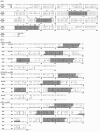
Fig. 1.
Fig. 1.
Sequence alignments of LANADBDs of gammaherpesviruses and EBNA1DBD reveals structural conservation. (a) Multiple alignments of amino acid sequences among LANADBDs of KSHV, RFHVMn and RRV using the praline and T-Coffee programs. Conserved amino acids among the OBPs are labelled in bold above the sequences. (b) Binary amino acid alignments between EBV EBNA1DBD and the LANADBDs of KSHV, RFHVMn and RRV using the 3d-pssm program. Conserved helices among the proteins are shown as shaded dark grey boxes. Proline loops are indicated in italic within light grey boxes. Numbers in parentheses refer to corresponding amino acid numbers from BC-1 KSHV LANA (Kelley-Clarke et al., 2007). Short dashes indicate missing amino acids; + and − indicate similarity or no similarity between amino acids.
Fig. 2.
Fig. 2.
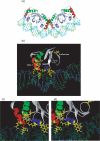
Computational model of the LANADBD and multimer structure bound to DNA. The figure shows the LANADBD model with the specific DNA-binding site predicted by the m-zdock program based on alignment with the structure of EBNA1DBD. (a) The tetramer formed by combining two dimers bound to their respective LBS1/2 (DNA helix in light blue and green). The β-barrel bundle is made of four β-strands from each monomer at the dimer interface. (b) Each monomer is composed of four β-strands and three helices (helix 1 in red, helix 2 in blue and helix 3 in green). (c, d) Crucial amino acids for DNA contact or dimerization are shown in yellow: 871K and 875Q for helix 1, 963W and 964E for helix 3 (c) and 907Y, 910K and 911K for helix 2 (d). The monomer pictures were generated using ViewerLite 4.2 (Accelrys).
Fig. 3.
Fig. 3.
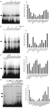
DNA-binding activity of LANADBD mutants. Purified LANADBD wt and mutant proteins were incubated with radiolabelled LBS1 or LBS1/2 as described previously (Garber et al., 2001). The DNA-binding affinity is represented as the percentage for mutants compared with wt LANADBD, which was set to 100 %. In each assay, all mutants were tested for DNA-binding activity with LBS1 (a–c) or LBS1/2 (d). EMSA results are shown for helix 1 mutants (a), helix 2 mutants (b), helix 3 mutants (c) and adapted mutants from each helix (d). Arrows indicate specific protein–DNA complexes. NC, Probe alone as a negative control; wt, wt LANADBD. Results on graphs are shown as means±sd from three independent experiments.
Fig. 4.
Fig. 4.
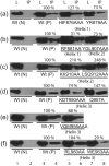
Co-immunoprecipitation assays with alanine substitution mutants. The dimerization ability of Flag-tagged wt or mutant LANADBDs with HA-tagged wt LANADBD was tested. Dimerization activity for each mutant was normalized based on the expression level of Flag-tagged wt or mutant LANADBD proteins. L, Cell lysate, IP; immunoprecipitated samples; Wt (N), HA-tagged wt only as a negative control; Wt (P), Flag-tagged and HA-tagged wt as a positive control.
Fig. 5.
Fig. 5.
Analysis of DNA replication mediated by alanine substitution mutants using a short-term replication assay. LANADBD-expressing constructs were co-transfected with pPuro/4TR into 293 cells; 10 % of the extracted DNA (Hirt, 1967) was digested with HindIII as input (a, lanes 1–8, and b, lanes 1–6) and the remaining DNA was double digested with HindIII and DpnI (a, lanes 9–16, and b, lanes 7–12). The DNA was detected by Southern blotting with a radiolabelled 4TR probe. Full-length LANA was transfected as a positive control. The arrow indicates the position of full-length pPuro-4TR.
Fig. 6.
Fig. 6.
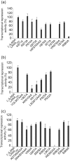
Analysis of the activity of LANA-dependent transcriptional repression by alanine substitution. Graphical representation of data from luciferase reporter assays. pGL3/7TR luciferase reporter and wt or mutant LANADBD plasmid were co-transfected. RLU values were normalized to total protein concentration as described previously (Renne et al., 2001). The percentage of suppression activity was compared with that of LANADBD wt, which was set to 100 %. Results are shown as means±sd from three independent experiments. (−)LANA, Negative control with no LANADBD.

Similar articles

Cited by

References

    1. Ballestas, M. E. & Kaye, K. M. (2001). Kaposi's sarcoma-associated herpesvirus latency-associated nuclear antigen 1 mediates episome persistence through cis-acting terminal repeat (TR) sequence and specifically binds TR DNA. J Virol 75, 3250–3258. - PMC - PubMed
    1. Ballestas, M. E., Chatis, P. A. & Kaye, K. M. (1999). Efficient persistence of extrachromosomal KSHV DNA mediated by latency-associated nuclear antigen. Science 284, 641–644. - PubMed
    1. Barbera, A. J., Chodaparambil, J. V., Kelley-Clarke, B., Joukov, V., Walter, J. C., Luger, K. & Kaye, K. M. (2006). The nucleosomal surface as a docking station for Kaposi's sarcoma herpesvirus LANA. Science 311, 856–861. - PubMed
    1. Bates, P. A., Kelley, L. A., MacCallum, R. M. & Sternberg, M. J. (2001). Enhancement of protein modeling by human intervention in applying the automatic programs 3d-jigsaw and 3d-pssm. Proteins (Suppl. 5), 39–46. - PubMed
    1. Bochkarev, A., Barwell, J. A., Pfuetzner, R. A., Furey, W., Jr, Edwards, A. M. & Frappier, L. (1995). Crystal structure of the DNA-binding domain of the Epstein–Barr virus origin-binding protein EBNA 1. Cell 83, 39–46. - PubMed

Publication types

MeSH terms

LinkOut - more resources