. 2010 Jul 15;21(14):2371-83.
doi: 10.1091/mbc.e10-02-0170. Epub 2010 May 19.

Aurora-C kinase deficiency causes cytokinesis failure in meiosis I and production of large polyploid oocytes in mice

Affiliations

Aurora-C kinase deficiency causes cytokinesis failure in meiosis I and production of large polyploid oocytes in mice

Kuo-Tai Yang et al. Mol Biol Cell. .

Abstract

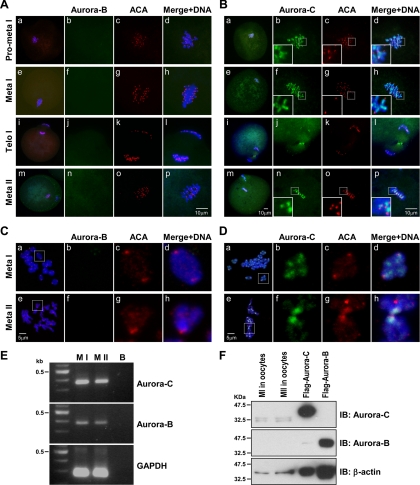
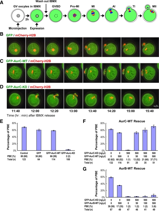
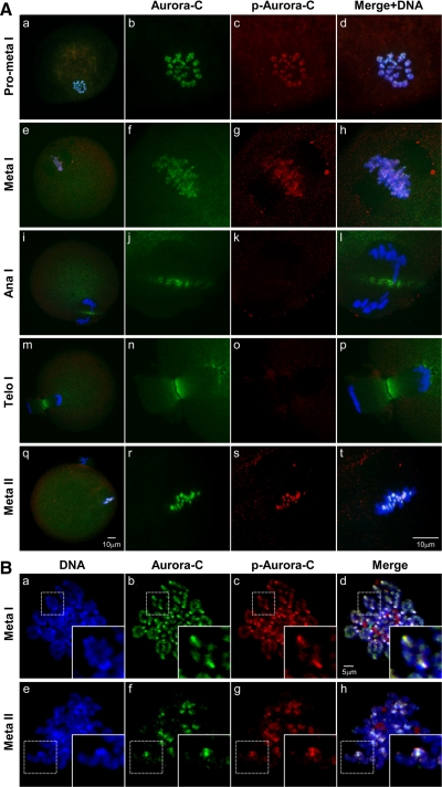
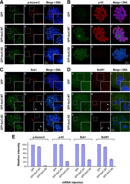
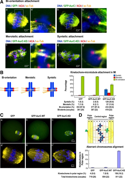
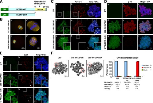
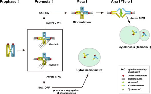
We previously isolated Aurora-C/Aie1 in a screen for kinases expressed in mouse sperm and eggs. Here, we show the localization of endogenous Aurora-C and examine its roles during female mouse meiosis. Aurora-C was detected at the centromeres and along the chromosome arms in prometaphase I-metaphase I and was concentrated at centromeres at metaphase II, in which Aurora-C also was phosphorylated at Thr171. During the anaphase I-telophase I transition, Aurora-C was dephosphorylated and relocalized to the midzone and midbody. Microinjection of the kinase-deficient Aurora-C (AurC-KD) mRNA into mouse oocytes significantly inhibited Aurora-C activity and caused multiple defects, including chromosome misalignment, abnormal kinetochore-microtubule attachment, premature chromosome segregation, and cytokinesis failure in meiosis I. Furthermore, AurC-KD reduced Aurora-C and histone H3 phosphorylation and inhibited kinetochore localization of Bub1 and BubR1. Similar effects also were observed in the oocytes injected with INCNEP-delIN mRNAs, in which the Aurora-C binding motif was removed. The most dramatic effect observed in AurC-KD-injected oocytes is cytokinesis failure in meiosis I, resulting in producing large polyploid oocytes, a pattern similar to Aurora-C deficiency human spermatozoa. Surprisingly, we detected no Aurora-B protein in mouse oocytes. We propose that Aurora-C, but not Aurora-B, plays essential roles in female mouse meiosis.


Figures

Figure 1.
Figure 1.
Expression and subcellular localization of Aurora-C, but not Aurora-B, in meiotic mouse oocytes. Confocal immunofluorescence analysis of endogenous Aurora-B (A and C) and Aurora-C (B and D) in whole-mount oocytes (A and B) or in chromosome spreads (C and D) during various stages of meiosis (prometaphase I ∼ meta II) by using indicated antibodies (green, Aurora-B and -C; red, ACA). ACA was used as a marker of the centromeric region of meiotic chromosomes. DNA was counterstained with DAPI (blue). In A and B, the image in A, b–d, and B, b–d (prometa I), is an enlarged view derived from Aa and Ba, respectively. Similar enlarged views are derived from the images in meta I (f–h from e), telo I (j–l from i), and meta II (n–p from m). In B, the large square box in b, c, d, f, g, h, n, o, and p is a blowup of each corresponding smaller dotted box. (C, b–d) An enlarged view of the boxed region derived from Ca; (C, f–h) region from Ce; (D, b–d) region from Da; (D, f–h) region from De. The Merge + DNA image is a triple merge of Aurora, ACA, and DNA. (E) Total RNAs isolated from MI and MII oocytes were analyzed by RT-PCR. Both Aurora-B and Aurora-C transcripts were detected in MI and MII oocytes. B, blank control. (F) Immunoblot analysis of the cell lysates prepared from mouse MI and MII oocytes (500 oocytes/lane) or from Flag-Aurora-B– (10 μg/lane) and Flag-Aurora-C (5 μg/lane)–transfected HeLa cells. Aurora-C was detected as doublet bands, possibly due to phosphorylation. No endogenous Aurora-B signal was detected in MI or MII oocytes.
Figure 2.
Figure 2.
The AurC-KD mutant induces early onset of chromosome segregation, failure to complete cytokinesis in meiosis I, and inhibition of first PBE. (A) Schematic representation of the process of microinjection during mouse oocyte maturation. Live mouse oocytes were coinjected mCherry-H2B (25 ng/μl; red) with each of GFP (500 ng/μl; B), GFP-AurC-WT (500 ng/μl; C), or GFP-AurC-KD (500 ng/μl; D) (green) mRNAs. The injection was performed in M2 medium containing 100 μM IBMX. The injected oocytes were cultured in IBMX-containing medium for 2 h. After incubation, the oocytes were cultured in IBMX-free medium for maturation. Photographs were taken with UltraView live cell imaging (LCI) confocal scanner (PerkinElmer Life and Analytical Sciences) at indicated times. Early onset of chromosome separation and cytokinesis failure were frequently observed in GFP-AurC-KD–injected oocytes (D). (E) Histogram showing percentages of first PBE in postinjected oocytes. Only postinjected oocytes with GVBD were calculated and counted. Number (n) of postinjected GVBD oocytes is shown. Grouped data were collected from three (GFP) to five (control, GFP-AurC-WT, and GFP-AurC-KD) independent experiments. Rescue of cytokinesis failure by AurC-WT (F) or AurB-WT (G) mRNA. GV oocytes were coinjected with a fixed concentration of GFP-AurC-KD transcripts (500 ng/μl) with increasing amounts of GFP-AurC-WT or GFP-AurB-WT mRNA (0–500 ng/μl). AurC-WT, but not AurB-WT, can rescue cytokinesis failure in GFP-AurC-KD–injected oocytes in a dose-dependent manner. Number (n) of postinjected GVBD oocytes is shown. Grouped data were collected from at least three independent experiments. Data are shown as mean ± SD.
Figure 3.
Figure 3.
Aurora-C deficiency induces premature chromosome segregation, resulting in univalent chromosomes in MI. (A) Chromosome spreads prepared from MI oocytes injected with GFP, GFP-AurC, or GFP-AurC-KD mRNAs 8 h post-GVBD. The oocytes were fixed and stained with DAPI as described in Materials and Methods. For a better view of chromosome morphology (bivalent vs. univalent), the DAPI image was converted to pseudocolor (black and white). (B) Histogram showing the percentage of bivalent (4N), univalent (2N), and singlet (1N) in oocytes injected with different mRNAs. Most chromosomes in GFP-AurC-KD–injected oocytes were univalent (∼97.3%). Chromatid number: n. Grouped data from four independent experiments.
Figure 4.
Figure 4.
Subcellular localization of endogenous Aurora-C and p-Aurora-C during meiotic divisions in mouse oocytes. Confocal immunofluorescence analysis of endogenous Aurora-C and p-Aurora-C in whole-mount oocytes (A) or in chromosome spreads (B) by using indicated antibodies (green for Aurora-C; red for p-Aurora-C). DNA was counterstained with DAPI (blue). In A, the image in A, b, c, and d (prometa I), is an enlarged view derived from Aa. Similar enlarged views are derived from the images in meta I (f–h from e), ana I (j–l from i), telo I (n–p from m), and meta II (r–t from q). The Merge + DNA image is a triple merge of Aurora-C, p-Aurora-C, and DNA. In B, the large square box in a–d (meta I) and e–h (meta II) is a blowup of each corresponding smaller dotted box.
Figure 5.
Figure 5.
AurC-KD inhibits kinetochore localization of Bub1 and BubR1 and reduces Aurora-C and histone H3 phosphorylation. The chromosome spreads (A–C) or whole-mount oocytes (D) were prepared either from prometaphase I oocytes (C and D; 6 h post-GVBD) or metaphase I oocytes (A and B; 8 h post-GVBD) injected with indicated mRNAs, followed by immunostaining with specific antibodies against phospho-Thr171-AurC (A), p-H3 (B), Bub1 (C), and BubR1 (D). DNA was counterstained with DAPI. The large square box in A, C, and D is a blowup of each corresponding smaller dotted box. The white arrow in C, e and h, and D, e, and h, indicates the position of kinetochore. The strong dotted p-Aurora-C signals (red) detected in A–H seem to be nonspecific, because these signals do not target to the centromeric regions. During normal oocyte maturation, Bub1 and BubR1 (spindle checkpoint proteins) signals were easily detected at the kinetochores on prometaphase I chromosomes (6 h post-GVBD) but frequently lost in metaphase-I chromosomes (8 h post-GVBD). The relative intensity of each injected mRNA compared with GFP (used as a control) is shown in E. Data are shown as mean ± SD.
Figure 6.
Figure 6.
AurC-KD induces aberrant K-MT attachment and chromosome misalignment in meiosis I. (A) Images were prepared from intact oocytes injected with GFP (a and a′, 8 h post-GVBD), GFP-AurC-WT (b and b′, 8 h post-GVBD), or GFP-AurC-KD (c and c′ and d and d′, 7 h post-GVBD) mRNAs. Under normal conditions, oocytes progress to metaphase I around 8 h after GVBD. Because, in GFP-AurC-KD–injected oocytes, the premature separation of bivalent chromosomes to univalent chromosomes was commonly occurred between 7 and 8 h post-GVBD, we thus examined the K-MT attachments in GFP-AurC-KD–injected oocytes at 7 h post-GVBD. The fixed oocytes were immunostained with antibodies against ACA and acetyl tubulin and analyzed by LSM510 laser confocal microscopy. The images shown in a–d are complied from five to eight confocal optical sections (0.8 μm/section). Enlarged views of each representative image (boxed) are shown on the right (a′–d′). The images shown in a′–d′ are complied from two optical sections (0.8 μm/section). (B) Statistical analysis of the percentage of aberrant kinetochore–microtubule attachments in postinjected oocytes. In GFP- and GFP-AurC-WT–injected oocytes, most kinetochore–microtubule attachments were in a biorientation arrangement, whereas merotelic and syntelic attachments were significantly increased in GFP-AurC-KD–injected oocytes. (C) The images were prepared as described in A. Most chromosomes were located at the central region in GFP- (C, a and a′) and GFP-AurC-WT (C, b and b′)–injected oocytes, whereas chromosome spread into the polar region was significantly increased in GFP-AurC-KD–injected oocytes (C, c and c′). (D, top) A cartoon showing the possible chromosome position during meiosis. (D, bottom) Statistical analysis of the percentage of aberrant chromosome alignment in postinjected oocytes. One bivalent carries two separated kinetochores. The pair of kinetochores on each sister chromatid fuses and functions as a unit during meiosis I. DNA was counterstained with DAPI. Grouped data were collected from at least three independent experiments. Data are shown as mean ± SD.
Figure 7.
Figure 7.
INCENP, like Aurora-C, is required for female meiotic divisions. (A) Schematic representation of a truncated form of INCENP (INCENP-delIN), in which the Aurora kinase binding motif (IN-Box) was removed. (B) Whole-mount images prepared from the MI oocytes were injected with indicated mRNAs and immunostained with anti-acetyl tubulin antibody. DNA was counterstained with DAPI (blue). (Ba′) An enlarged view of chromosomes derived from Ba; (Bb′) that derived from Bb. (C–E) Chromosome spreads prepared from the oocytes injected with indicated mRNAs were immunostained with anti-Aurora-C (C; 8 h post-GVBD), anti-phospho-H3 (D; 8 h post-GVBD), or anti-Bub1 antibodies (E; 6 h post-GVBD) and analyzed by LSM510 laser confocal microscopy. The large square box in C and E is a blowup of each corresponding smaller dotted box. The white arrow in Ee and Eh indicates the position of kinetochore. (F) Left, chromosome spreads prepared from the MI oocytes (8 h post-GVBD) injected with indicated mRNAs were fixed and stained with DAPI. For a better view of chromosome morphology (bivalent vs. univalent) in F, the DAPI image was converted to pseudocolor (black and white). Right, percentage of bivalent (4N), univalent (2N), and singlet (1N) in oocytes injected with different mRNAs. Most chromosomes in GFP-INCENP-delIN–injected oocytes were univalents (97%). Bottom, grouped data from four independent experiments. Chromatid number, n.
Figure 8.
Figure 8.
Schematic representation of the subcellular localization of Aurora-C and its possible functions in female meiosis. During late prophase I–metaphase I, Aurora-C is phosphorylated at Thr171 and located at the centromeric regions as well as along the chromosome arms (see Figure 4). Aurora-C is dephosphorylated and relocalized to the midzone and midbody (see Figure 4) during anaphase I–telophase I transition and then rephosphorylated and concentrated again to the centromeres at metaphase II (see Figure 1, B and D). During prometaphase-metaphase I, Aurora-C is involved in promoting chromosome biorientation by correcting aberrant kinetochore-microtubule attachments in meiosis I, whereas its deficiency causes early onset of chromosome segregation in MI and failure to complete cytokinesis. Aurora-C-KD, Aurora-C kinase-deficient mutant.

References

    1. Andrews P. D., Knatko E., Moore W. J., Swedlow J. R. Mitotic mechanics: the auroras come into view. Curr. Opin. Cell Biol. 2003;15:672–683. - PubMed
    1. Bernard M., Sanseau P., Henry C., Couturier A., Prigent C. Cloning of STK13, a third human protein kinase related to Drosophila aurora and budding yeast Ipl1 that maps on chromosome 19q13.3-ter. Genomics. 1998;53:406–409. - PubMed
    1. Carmena M., Earnshaw W. C. The cellular geography of aurora kinases. Nat. Rev. Mol. Cell Biol. 2003;4:842–854. - PubMed
    1. Chang C. J., Goulding S., Adams R. R., Earnshaw W. C., Carmena M. Drosophila Incenp is required for cytokinesis and asymmetric cell division during development of the nervous system. J. Cell Sci. 2006;119:1144–1153. - PMC - PubMed
    1. Chen H. L., Tang C. J., Chen C. Y., Tang T. K. Overexpression of an Aurora-C kinase-deficient mutant disrupts the Aurora-B/INCENP complex and induces polyploidy. J. Biomed. Sci. 2005;12:297–310. - PubMed

Publication types

MeSH terms

LinkOut - more resources