. 2010 Jul 15;21(14):2453-68.
doi: 10.1091/mbc.e09-12-1031. Epub 2010 May 19.

Lens fiber cell differentiation and denucleation are disrupted through expression of the N-terminal nuclear receptor box of NCOA6 and result in p53-dependent and p53-independent apoptosis

Affiliations

Lens fiber cell differentiation and denucleation are disrupted through expression of the N-terminal nuclear receptor box of NCOA6 and result in p53-dependent and p53-independent apoptosis

Wei-Lin Wang et al. Mol Biol Cell. .

Abstract

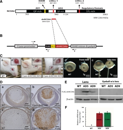
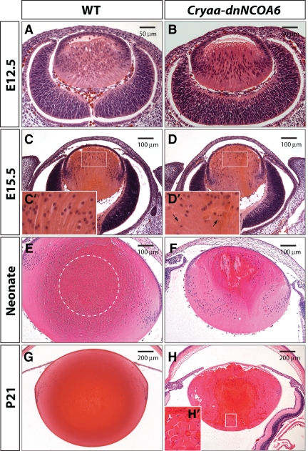
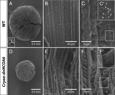
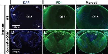
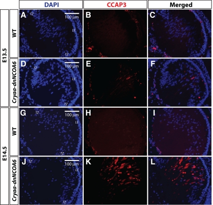
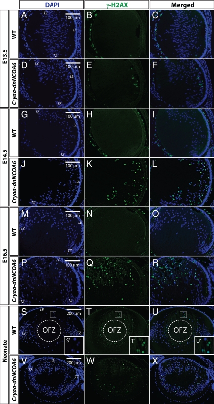
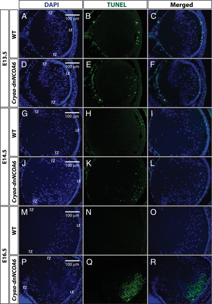
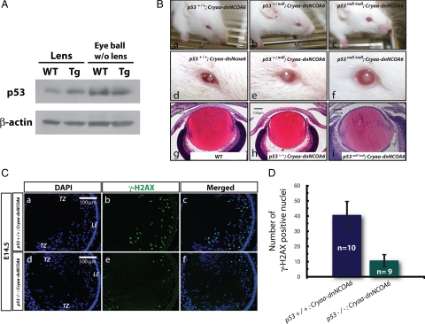
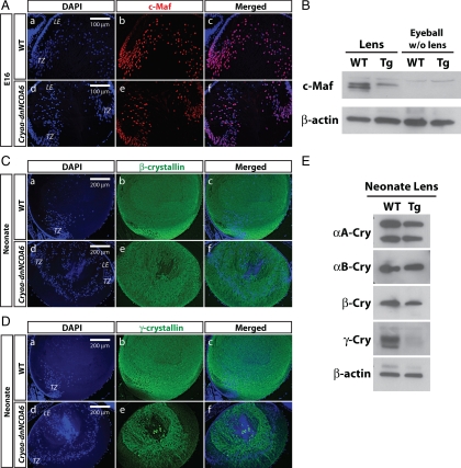
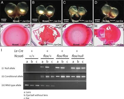
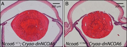
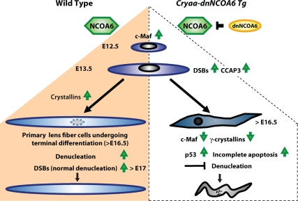
Nuclear receptor coactivator 6 (NCOA6) is a multifunctional protein implicated in embryonic development, cell survival, and homeostasis. An 81-amino acid fragment, dnNCOA6, containing the N-terminal nuclear receptor box (LXXLL motif) of NCOA6, acts as a dominant-negative (dn) inhibitor of NCOA6. Here, we expressed dnNCOA6 in postmitotic transgenic mouse lens fiber cells. The transgenic lenses showed reduced growth; a wide spectrum of lens fiber cell differentiation defects, including reduced expression of gamma-crystallins; and cataract formation. Those lens fiber cells entered an alternate proapoptotic pathway, and the denucleation (karyolysis) process was stalled. Activation of caspase-3 at embryonic day (E)13.5 was followed by double-strand breaks (DSBs) formation monitored via a biomarker, gamma-H2AX. Intense terminal deoxynucleotidyl transferase dUTP nick-end labeling (TUNEL) signals were found at E16.5. Thus, a window of approximately 72 h between these events suggested prolonged though incomplete apoptosis in the lens fiber cell compartment that preserved nuclei in its cells. Genetic experiments showed that the apoptotic-like processes in the transgenic lens were both p53-dependent and p53-independent. Lens-specific deletion of Ncoa6 also resulted in disrupted lens fiber cell differentiation. Our data demonstrate a cell-autonomous role of Ncoa6 in lens fiber cell differentiation and suggest novel insights into the process of lens fiber cell denucleation and apoptosis.


Figures

Figure 1.
Figure 1.
Generation and phenotype analysis of the Cryaa-dnNCOA6 transgenic mouse model. (A) Schematic diagram of the NCOA6 protein structure (2063 amino acid residues). NCOA6 contains two QP-rich ADs, two canonical NR boxes (LXXLL-1 and LXXLL-2), which are important for interaction with ligand-bound NRs, one noncanonical NR box (IXXMM), and the C-terminal STL-rich regulatory domain. (B) Schematic diagram of the Cryaa-dnNCOA6 transgenic construct. cDNA of dnNCOA6 including amino acid 849–929 of NCOA6 with a NLS and 3xFLAG tag in the 5′ end was inserted between the Cryaa (αA-crystallin) promoter and the intron-polyadenylation sequences of the small t antigen from SV40 (SV40 intron & polyA). The three primers used for genotyping are shown as horizontal arrows. (C) Two founders of Cryaa-dnNCOA6, AD5 (b) and AD9 (c), were generated and showed microphthalmia and cataract compared with WT (a) mice. Eyeballs from WT and AD5 animals are aligned in front view (d) and side view (e) to compare phenotypes. Transgenic lenses displayed a reduction of ∼3.84- to 4.75-fold in size compared with WT (see Materials and Methods). Transgene copy number of line AD5 and AD9 is 12∼16 and 12∼19 copies per genome, respectively. (D) Lens-specific expression of the FLAG-dnNCOA6 fragment was evaluated by immunohistochemistry using an anti-FLAG antibody (brown) in E14.5 WT (a and c) and transgenic (b and d) eyes. Nuclei (blue) were counterstained with hematoxylin. Scale bar is shown in each panel. (E) Western blot analysis of lens-specific FLAG-dnNCOA6 fragments expression in the lens and eyeball (without lens) tissues from WT, AD5, and AD9. β-Actin was used for loading control. (F) Relative expression levels of transcripts encoding dnNCOA6 in AD5 and AD9 transgenic lines were analyzed by qRT-PCR. Primers used (see Materials and Methods) are specific to the transgene (human sequence) and did not cross-react with the endogenous mouse Ncoa6 transcripts.
Figure 2.
Figure 2.
Histological analysis of lens development revealed lens fiber cell differentiation defects in Cryaa-dnNCOA6 lenses. H&E staining was performed with WT (A, C, E, and G) and transgenic (B, D, F, and H) mouse eye sections at E12.5, E15.5, neonatal stage, and P21, respectively, to show lens morphology. At E15.5, the transgenic lens appeared smaller (D) compared with WT (C) and exhibits pyknotic nuclei. Higher magnification images of E15.5 lens fiber cell nuclei are shown as insets (C′ and D′) and pyknotic nuclei are indicated by arrows. The OFZ is located in the center of the WT lens (E and G) and is indicated by dashed circle in E. Transgenic lenses display irregular fiber pattern and pyknotic staining (F and H). The rounded end fragment and cortical liquefaction in dark red are revealed in the inset (H′). Scale bar is shown in each panel.
Figure 3.
Figure 3.
Loss of Y-suture and deformed lens fiber cells in Cryaa-dnNCOA6 lenses. SEM analysis was carried out on 3-mo-old lenses from WT (A–C) and transgenic mice (D–F). Normal Y-suture found in the WT lens (A) is absent in the transgenic lens (D). The Y-suture is illustrated as inset in A. Higher magnification reveals the enlarged and disrupted fiber cell pattern in the transgenic lens (E), whereas the WT lens showed aligned fiber cell pattern (B). Ball-and-socket structure (indicated by white arrows in C′), which is important to hold fiber cells together, is not apparent in the transgenic lens (F and F′) compared with the WT lens (C and C′). Scale bar is given in each panel.
Figure 4.
Figure 4.
OFZ is absent in the Cryaa-dnNCOA6 lens. An anti-PDI antibody was used to label ER, indicating the existence of organelles, in WT (A–C) and transgenic lenses (D–F) at neonatal stage. Nuclei were stained (blue signal) with DAPI. The center of WT lens cortex is negative with DAPI and anti-PDI antibody staining, which is labeled as OFZ. Lens epithelium, LE; transitional zone (bow region), TZ. Scale bar is shown in A and D.
Figure 5.
Figure 5.
Caspase-3 activation in differentiating transgenic lens fiber cells. E13.5 and E14.5 WT and transgenic lenses were subjected to immunofluorescence analysis with CCAP3 antibody. The E13.5 transgenic lens showed staining in the lens fiber cell compartment (D–F) compared with WT (A–C). The amount of CCAP3 staining increased in the E14.5 transgenic lens (J–L) and no staining was detected in the WT lens (G–I). Nuclei (blue signal) were stained with DAPI. Lens epithelium, LE; transitional zone, TZ. Scale bar is shown in panels A, D, G, and J.
Figure 6.
Figure 6.
Identification of DNA double-strand breaks (DSBs) through γ-H2AX immunofluorescence in WT and Cryaa-dnNCOA6 lens fiber cells. γ-H2AX antibody was used to label DSBs generated through DNA damage or normal denucleation process. DSBs are detected as early as E13.5 in the transgenic lens fiber cell compartment (D–F) and are absent in the WT lens (A–C). The number of DSBs increased in E14.5 and E16.5 transgenic lens fiber cells (J–L and P–R), whereas WT controls are negative (G–I and M–O). In the neonatal WT lens, nuclei of lens fiber cells at the margin of the OFZ display strong γ-H2AX staining (S–U and S′–U′). The neonatal transgenic lens exhibits dispersed DSBs staining in fiber cells and the absence of OFZ (V–X). The OFZ is indicated by dashed circle. Nuclei (blue signal) were stained with DAPI. Mesenchymal cells and hyaloid vascular structure surrounding the lens often generate nonspecific signals, which do not interfere with our observations in the lens fiber cell compartment. Lens epithelium, LE; transitional zone, TZ. Scale bar is shown in A, D, G, J, M, P, S, and V.
Figure 7.
Figure 7.
TUNEL assays in WT and Cryaa-dnNCOA6 lens fiber cells. (A–C) At E13.5, sporadic apoptotic nuclei were detected only in the epithelium cells of WT lenses. (D–F) In contrast, apoptotic nuclei were detected in both epithelium and lens fiber cells of transgenic lenses. TUNEL-positive nuclei: 5.47 ± 2.00 nuclei/section. (J–L) Similar results were observed in E14.5 lenses with slightly elevated number of apoptotic nuclei in transgenic lens fiber cells. TUNEL-positive nuclei: 12.88 ± 7.59 nuclei/section. (P–R) At E16.5, strong TUNEL signals were detected in the frontal part of lens fiber cell compartment in the transgenic lens. No TUNEL-positive nuclei were observed in WT lens fiber cells (A–C, G–I, and M–O). Nonspecific signals outside of the lens are explained in Figure 6. Lens epithelium, LE; transitional zone, TZ. Scale bar is shown in A, D, G, J, M, and P.
Figure 8.
Figure 8.
Abnormal lens fiber cell apoptosis is both p53-dependent and p53-independent. (A) Western blot was performed to analyze p53 protein expression in the lens and eyeball (without lens) of WT and transgenic mice at neonatal stage. β-Actin was used for loading control. (B) External observation of adult mouse eyes (a–f) and histological analysis of E16.5 mouse eyes (g–i) in p53+/+; Cryaa-dnNCOA6, p53+/null; Cryaa-dnNCOA6, p53null/null; Cryaa-dnNCOA6, and WT mice. (C) Reduced number of DSBs in p53null/null; Cryaa-dnNCOA6 lenses (d–f) compared with p53+/+; Cryaa-dnNCOA6 lenses at E14.5 (a–c). Nuclei (blue signal) were stained with DAPI. Scale bar is shown in a and d. (D) To quantify the amounts of DSBs in Cryaa-dnNCOA6 lenses in WT or p53 null background, the number of γ-H2AX-positive nuclei was compared between E14.5 p53+/+; Cryaa-dnNCOA6 (n = 10) and p53null/null; Cryaa-dnNCOA6 (n = 9) lenses. Nonspecific signals outside of the lens are explained in Figure 6. Lens epithelium, LE; transitional zone, TZ.
Figure 9.
Figure 9.
Down-regulation of c-Maf and γ-crystallins in the Cryaa-dnNCOA6 transgenic lenses. (A) Immunofluorescence staining was carried out in E16 WT and transgenic lenses. In WT, c-Maf protein expression was up-regulated in the transitional zone and persisted in lens fiber nuclei (A, a–c). However, many lens fiber nuclei lost c-Maf protein expression in the transgenic lens (A, d–f). Nuclei (blue signal) were stained with DAPI. (B) Western blot analysis of the c-Maf protein expression level in WT and transgenic neonatal lenses (2.37- to 2.64-fold reduction in transgenic lenses) and eyeballs (without lens). β-Actin was used as loading control. (C) Some reduction of β-crystallins in transgenic lenses was observed compared with WT lenses. Lens epithelium, LE; transitional zone, TZ. (D) γ-Crystallin proteins expression is down-regulated in transgenic lenses compared with WT lenses. (E) Western blot analysis of αA-crystallin (αA-Cry), αB-crystallin (αB-Cry), β-crystallin (β-Cry), and γ-crystallin (γ-Cry) protein expression levels in neonatal WT and transgenic lenses. β-Actin served as loading control. The protein expression ratios of WT to transgenic lenses are indicated as follows: αA-Cry (1.08–1.18), αB-Cry (1.18–1.23), β-Cry (1.46–1.56), and γ-Cry (1.56–2.95).
Figure 10.
Figure 10.
Deletion of Ncoa6 in prospective lens ectoderm led to microphthalmia and cataract. (A–D) Eyeball morphology of four genotypes: Ncoa6+/+; Le-Cre, Ncoa6flox/+; Le-Cre, Ncoa6flox/flox; Le-Cre, and Ncoa6flox/null; Le-Cre at ∼3 mo old. (E–H) H&E staining of 3-mo-old lenses from Ncoa6+/+; Le-Cre and Ncoa6flox/flox; Le-Cre mice, and 1-mo-old lenses from Ncoa6+/null; Le-Cre and Ncoa6flox/null; Le-Cre mice. Scale bar is shown in each panel. (I) To analyze the deletion efficiency of the floxed allele, lens, eyeball (without lens), and ear tissues from Ncoa6+/+; Le-Cre, Ncoa6flox/+; Le-Cre, Ncoa6flox/flox; Le-Cre, and Ncoa6flox/null; Le-Cre mice were used to extract genomic DNA for genotyping. Analysis of ear tissue confirmed the tissue-specific expression of Le-Cre. In Ncoa6+/+; Le-Cre tissues, only the WT allele was detected. Most Ncoa6flox/+; Le-Cre tissues showed the WT, conditional and null alleles except ear tissues in which no null allele is detected. In Ncoa6flox/flox; Le-Cre mice, the null allele is detected in the lens; however, the deletion is incomplete as in the Ncoa6flox/null; Le-Cre lens.
Figure 11.
Figure 11.
Increased lens abnormalities in Ncoa6+/null; Cryaa-dnNCOA6 mice. Heterozygous Ncoa6 mice were crossed with Cryaa-dnNCOA6 mice to generate Ncoa6+/+; Cryaa-dnNCOA6 and Ncoa6 +/null; Cryaa-dnNCOA6 mice. Serial eye sections were used to compare the lens size of each genotype. The result indicates that the Ncoa6+/null; Cryaa-dnNCOA6 lens (B) is smaller than the Ncoa6+/+; Cryaa-dnNCOA6 lens (A). Quantitative analysis using P21 lenses revealed that the Ncoa6 +/null; Cryaa-dnNCOA6 lens is 23 ± 11% smaller than the Ncoa6+/+; Cryaa-dnNCOA6 lens. Eyes from three mice of each genotype were analyzed. Scale bar is shown in each panel.
Figure 12.
Figure 12.
Summary diagram of lens fiber cell differentiation and the role of NCOA6 in the denucleation process. In WT lens (left side of the diagram), up-regulation of c-Maf and crystallins is essential for lens fiber cell differentiation. Their terminal differentiation requires orchestrated degradation of all subcellular organelles to avoid light scattering. Nuclear degradation is a relatively lengthy process (3–4 d in mouse; Bassnett, 2009). In Cryaa-dnNCOA6 transgenic lenses (right side of the diagram), p53-dependent and p53-independent proapoptotic processes become evident as early as at E13.5 due to the generation of CCAP3 followed by the appearance of DSBs monitored via γ-H2AX–specific antibodies. Although abundant TUNEL signals are found at E16.5, the nuclear degradation is not properly executed and abnormally differentiated lens fibers, with significantly reduced expression of c-Maf and γ-crystallins and retained nuclei, are preserved in the transgenic lenses.

Similar articles

Cited by

References

    1. Alge C. S., Priglinger S. G., Neubauer A. S., Kampik A., Zillig M., Bloemendal H., Welge-Lussen U. Retinal pigment epithelium is protected against apoptosis by alphaB-crystallin. Invest. Ophthalmol. Vis. Sci. 2002;43:3575–3582. - PubMed
    1. Andley U. P. Crystallins in the eye: function and pathology. Prog. Retin. Eye Res. 2007;26:78–98. - PubMed
    1. Antonson P., Schuster G. U., Wang L., Rozell B., Holter E., Flodby P., Treuter E., Holmgren L., Gustafsson J. A. Inactivation of the nuclear receptor coactivator RAP250 in mice results in placental vascular dysfunction. Mol. Cell Biol. 2003;23:1260–1268. - PMC - PubMed
    1. Anzick S. L., Kononen J., Walker R. L., Azorsa D. O., Tanner M. M., Guan X. Y., Sauter G., Kallioniemi O. P., Trent J. M., Meltzer P. S. AIB1, a steroid receptor coactivator amplified in breast and ovarian cancer. Science. 1997;277:965–968. - PubMed
    1. Ashery-Padan R., Marquardt T., Zhou X., Gruss P. Pax6 activity in the lens primordium is required for lens formation and for correct placement of a single retina in the eye. Genes Dev. 2000;14:2701–2711. - PMC - PubMed

Publication types

MeSH terms