. 2010 May 19;30(20):6852-61.
doi: 10.1523/JNEUROSCI.0761-10.2010.

The neurotoxic effects of estrogen on ischemic stroke in older female rats is associated with age-dependent loss of insulin-like growth factor-1

Affiliations

The neurotoxic effects of estrogen on ischemic stroke in older female rats is associated with age-dependent loss of insulin-like growth factor-1

Amutha Selvamani et al. J Neurosci. .

Abstract

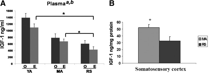
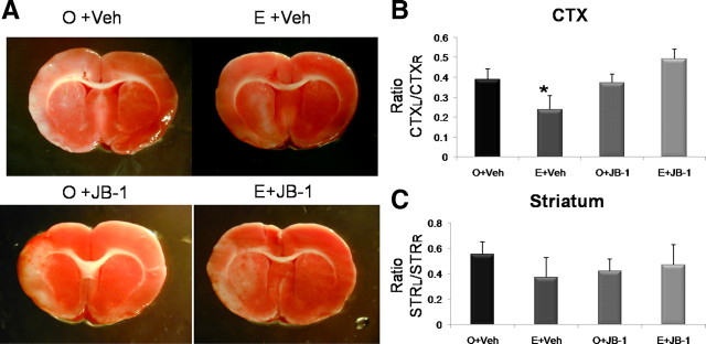
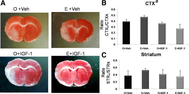
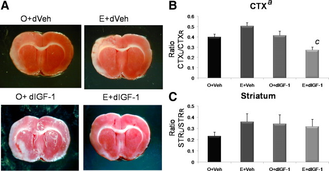
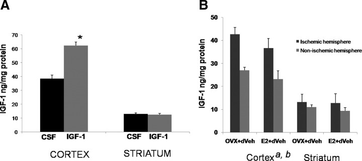
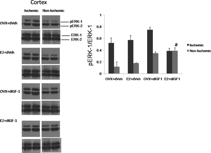
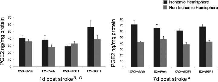
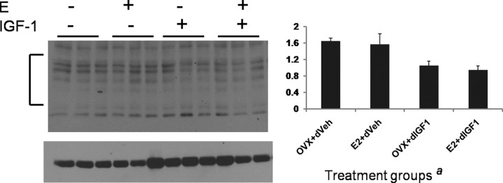
Hormone therapy to postmenopausal females increases the risk and severity of ischemic stroke. Our previous work using an animal model of menopause (reproductive senescence) shows that middle cerebral artery occlusion (MCAo) causes a larger cortical-striatal infarct in this older acyclic group compared with younger females. Moreover, although estrogen treatment is neuroprotective in younger females, estrogen paradoxically increases infarct volume in acyclic females. We hypothesized that the neurotoxic effects of estrogen in older females occurs because of decreased availability of IGF-1, a neuroprotectant that decreases with advancing age and is downregulated by estrogen treatment. Our data show that plasma IGF-1 levels are significantly reduced in reproductive senescent females and further reduced by estrogen at all ages. The neuroprotective effect of estrogen on MCAo-induced cortical infarct volume in mature adult female is reversed by intracerebroventricular injections of IGF-1 receptor antagonist JB-1. Similarly, estrogens neurotoxic effects on cortical infarct volume in senescent females is attenuated by concurrent IGF-1 treatment, and reversed when IGF-1 is infused 4 h after the onset of ischemia (delayed IGF-1 treatment). Delayed IGF-1/estrogen treatment also suppressed ischemia-induced ERK1 phosphorylation, reduced protein oxidation, and stimulated an early increase in prostaglandin E(2) at the infarct site. IGF-1 treatment was only protective in senescent females that received estrogen, indicating that the neuroprotective actions of this peptide require interaction with the steroid hormone receptor. These data support the hypothesis that stroke severity in older females is associated with decreased IGF-1 and further indicate that short-term postischemic IGF-1 therapy may be beneficial for stroke.


Figures

Figure 1.
Figure 1.
Circulating and local levels of IGF-1. A, IGF-1 levels in plasma of young adults, mature adults, and reproductive senescent females were determined by ELISA. Both age (a, main effect of age, p < 0.05) and estrogen treatment (b, main effect of hormone, p < 0.05) significantly reduced IGF-1 levels. Planned comparisons indicate that IGF-1 levels in the reproductive senescent females are significantly lower than those in the young adult and mature adults. B, IGF-1 levels in somatosensory cortex. Mature adult females had significantly higher levels of IGF-1 compared with reproductive senescent females (*p ≤ 0.05). YA, Young adults (3–4 months of age); MA, mature adult females (5–6 months); RS, reproductive senescent females (9–11 months). Error bars indicate SEM.
Figure 2.
Figure 2.
Effects of estrogen and concurrent JB-1 treatment on infarct size in mature adult females. MCAo induced by ET-1 injections resulted in cortical and striatal lesions as indicated by TTC-stained sections (A). The volume of the cortical infarct was significantly reduced in the estrogen-treated mature adult females (E) compared with the control-replaced females (O), whereas JB-1 treatment reversed this effect. Neither estrogen nor JB-1 affected striatal infarct volume. Histogram depicts mean ± SEM of cortical (B) and striatal (C) infarct volume as a ratio of the contralateral cortex and striatum, respectively. n = 4–6 in each group; *p < 0.05.
Figure 3.
Figure 3.
Effects of estrogen and concurrent IGF-1 treatment on infarct size in reproductive senescent females. MCAo induced by ET-1 injections resulted in cortical and striatal infarcts, as indicated by the TTC staining (A). The volume of the cortical infarct was significantly greater in the estrogen-treated reproductive senescent females (E) compared with the control-replaced females (O). Intracerebroventricular infusion of IGF-1 concurrent with the onset of ischemia attenuated infarct size (a, main effect of IGF-1, p < 0.05). Planned comparisons indicate that cortical infarct size is reduced in the E+IGF-1 group compared with the E+Veh, but is equivalent to the O+Veh group. Neither estrogen nor IGF-1 had any effect on striatal infarct volume. Histogram depicts mean ± SEM of cortical (B) and striatal (C) infarct volume as a ratio of the contralateral cortex and striatum, respectively. n = 4–6 in each group.
Figure 4.
Figure 4.
Effect of estrogen and delayed IGF-1 treatment on infarct size in reproductive senescent females. A, TTC-stained sections from the brain of animals subject to ischemic stroke induced by ET-1 injections. Cortical infarct volume was significantly greater in estrogen-treated reproductive senescent females (E) compared with the control-replaced females (O). Intracerebroventricular infusion of IGF-1 4 h after ischemia (delayed IGF-1) reduced cortical infarct volume (a, main effect of IGF-1, p < 0.05). There was a significant interaction effect of E+IGF-1 (c, interaction effect, p < 0.05). Planned comparisons indicate that the cortical infarct volume was significantly reduced in the E+IGF-1 group compared with all other groups including E+Veh and the O+Veh. No effect of estrogen or delayed IGF-1 was seen on striatal infarct volume. B, C, Histogram depicts mean ± SEM of cortical (B) and striatal (C) infarct volume as a ratio of the contralateral cortex and striatum, respectively. n = 4–7 in each group.
Figure 5.
Figure 5.
IGF-1 expression in the cortex and striatum of reproductive senescent females. A, IGF-1 was infused 4 h after the onset of ischemia and was measured 24 h later. Intracerebroventricular infusions of IGF-1 significantly increased peptide accumulation in the cortex, but not in the striatum. B, Endogenous levels of IGF-1 levels were increased in the ischemic cortex compared with the nonischemic (right) hemisphere (a, main effect of hemisphere, repeated measure, p < 0.05), whereas estrogen-treated animals had lower levels of IGF-1 (b, main effect of estrogen, p < 0.05). No significant differences were observed in the striatum. *p < 0.05. Error bars indicate SEM.
Figure 6.
Figure 6.
Western blot analysis of phospho- and pan-ERK expression in cortical lysates from the ischemic and nonischemic hemispheres of reproductive senescent females. Although pERK-1 expression (normalized to ERK-1) was elevated in the ischemic hemisphere in most treatment groups, estrogen treatment combined with delayed IGF-1 infusion suppressed ischemia-induced ERK-1 activation (a, interaction effect of hemisphere, estrogen, IGF-1). n = 3–4 per group. Error bars indicate SEM.
Figure 7.
Figure 7.
PGE2 expression in the ischemic and nonischemic cortex. PGE2 levels were measured by ELISA and normalized to total protein. At 24 h after MCAo, PGE2 levels were modestly elevated in the ischemic cortex (a, main effect of hemisphere, repeated measure, p < 0.05). The levels of PGE2 were highest in the group that received estrogen and IGF-1 (c, interaction effect of E+IGF-1, p < 0.05). At 7 d after MCAo, PGE2 levels were elevated in the ischemic cortex of all groups (p < 0.05, main effect of hemisphere, repeated measure) to a similar extent. n = 4–6 per group. Error bars indicate SEM.
Figure 8.
Figure 8.
Western blot analysis of 2,4-DNP-modified proteins in ischemic cortical lysates from senescent animals treated with estrogen or control pellet and later treated intracerebroventricularly with either IGF-1 or vehicle 4 h after ischemia. Proteins were derivatized by incubating tissue lysates with 2,4-dinitrophenylhydrazine. Immunosignal for 2,4-DNP proteins normalized to a loading control (tubulin) was significantly decreased in groups that received IGF-1 infusion (a, main effect of IGF-1, p < 0.05). The bracket indicates the region in each lane that was quantitated by densitometry. n = 3 per group. Error bars indicate SEM.
Figure 9.
Figure 9.
ERα expression and immunoprecipitation with IGF-1R in the cortex of reproductive senescent females. A, Western blot assays showing ERα expression in both the ischemic and nonischemic cortex. In the nonischemic cortex, ERα expression, normalized to tubulin, was similar in all groups. In the ischemic hemisphere, ERα expression was significantly higher in groups that received IGF-1 (c, interaction effect of hemisphere and IGF-1, p < 0.05). Error bars indicate SEM. B, ERα/IGF-1R immunoprecipitation. Cortical lysates immunoprecipitated with ERα antibody revealed IGF-1R b segment (78 kDa) immunosignal (i), and parallel experiments of cortical lysates immunoprecipitated with IGF-1R revealed ERα (68 kDa) immunosignal (ii). ERα /IGF1R complexes were seen in all treatments; shown here are representative samples from the reproductive senescent animals treated with an estrogen pellet and intracerebroventricular infusion of IGF-1.

Similar articles

Cited by

References

    1. Alkayed NJ, Murphy SJ, Traystman RJ, Hurn PD, Miller VM. Neuroprotective effects of female gonadal steroids in reproductively senescent female rats. Stroke. 2000;31:161–168. - PubMed
    1. Bartke A, Chandrashekar V, Dominici F, Turyn D, Kinney B, Steger R, Kopchick JJ. Insulin-like growth factor 1 (IGF-1) and aging: controversies and new insights. Biogerontology. 2003;4:1–8. - PubMed
    1. Biernaskie J, Corbett D, Peeling J, Wells J, Lei H. A serial MR study of cerebral blood flow changes and lesion development following endothelin-1-induced ischemia in rats. Magn Reson Med. 2001;46:827–830. - PubMed
    1. Böttner M, Wuttke W. Chronic treatment with physiological doses of estradiol affects the GH-IGF-1 axis and fat metabolism in young and middle-aged ovariectomized rats. Biogerontology. 2006;7:91–100. - PubMed
    1. Brown-Borg HM. Hormonal regulation of longevity in mammals. Ageing Res Rev. 2007;6:28–45. - PMC - PubMed

Publication types

MeSH terms