. 2010 May 19;30(20):6862-72.
doi: 10.1523/JNEUROSCI.1051-10.2010.

Liver X receptor agonist treatment ameliorates amyloid pathology and memory deficits caused by high-fat diet in APP23 mice

Affiliations

Liver X receptor agonist treatment ameliorates amyloid pathology and memory deficits caused by high-fat diet in APP23 mice

Nicholas F Fitz et al. J Neurosci. .

Abstract

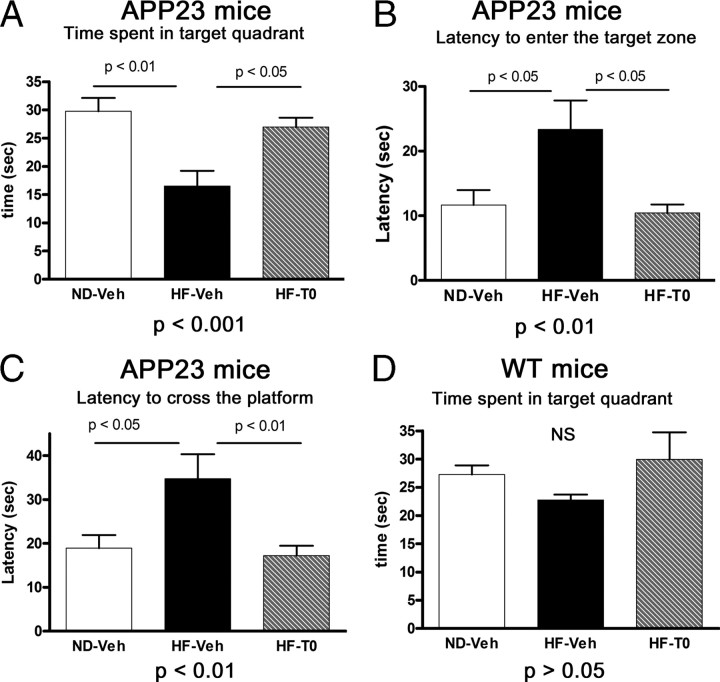
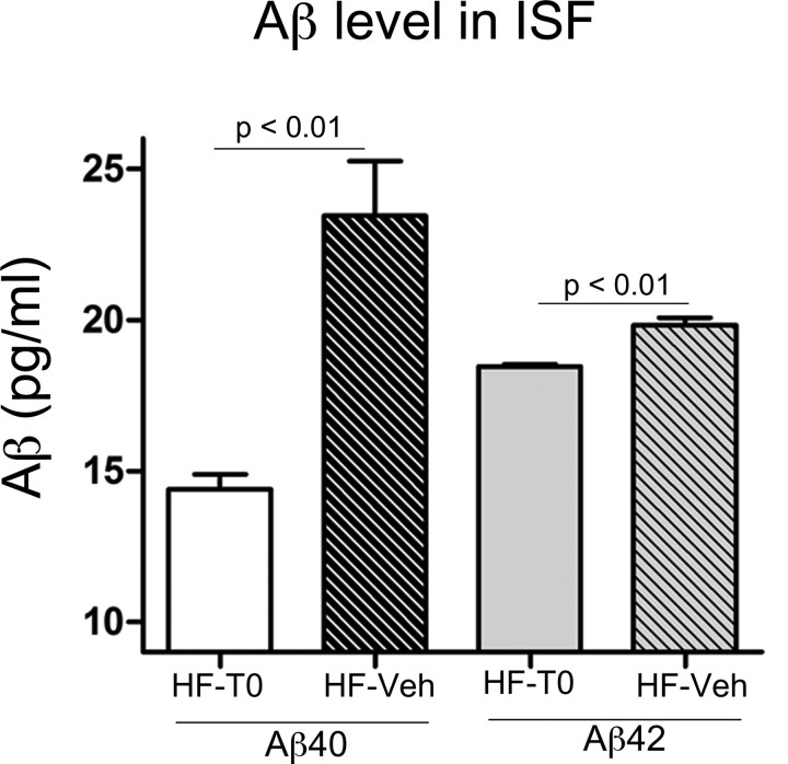
High-fat diet and certain dietary patterns are associated with higher incidence of sporadic Alzheimer's disease (AD) and cognitive decline. However, no specific therapy has been suggested to ameliorate the negative effects of high fat/high cholesterol levels on cognition and amyloid pathology. Here we show that in 9-month-old APP23 mice, a high-fat/high-cholesterol (HF) diet provided for 4 months exacerbates the AD phenotype evaluated by behavioral, morphological, and biochemical assays. To examine the therapeutic potential of liver X receptor (LXR) ligands, APP23 mice were fed HF diet supplemented with synthetic LXR agonist T0901317 (T0). Our results demonstrate that LXR ligand treatment causes a significant reduction of memory deficits observed during both acquisition and retention phases of the Morris water maze. Moreover, the effects of T0 on cognition correlate with AD-like morphological and biochemical parameters. We found a significant decrease in amyloid plaque load, insoluble Abeta and soluble Abeta oligomers. In vitro experiments with primary glia demonstrate that Abca1 is essential for the proper lipidation of ApoE and mediates the effects of T0 on Abeta degradation by microglia. Microdialysis experiments performed on awake freely moving mice showed that T0 decreased Abeta levels in the interstitial fluid of the hippocampus, supporting the conclusion that this treatment increases Abeta clearance. The data presented conclusively shows that LXR activation in the context of a metabolic challenge has critical effects on AD phenotype progression by attenuating Abeta deposition and facilitating its clearance.


Figures

Figure 1.
Figure 1.
High-fat diet aggravates spatial learning impairment in APP23 mice and T0901317 ameliorates the deficit: MWM acquisition test. A, Path length scores represent the average distance swum to find the platform per trial block. (1) There was a significant treatment effect on path length (F(2,41) = 10.52, p < 0.0001); (2) a treatment × trial block interaction significantly affects path length as well (F(8,164) = 2.59, p < 0.01). p < 0.05, ND-Veh compared to HF-Veh by Tukey's post hoc test. B, Escape latency scores represent the average acquisition time to find the platform per trial block. (1) There was a significant treatment effect on escape latency (F(2,41) = 18.34, p < 0.0001); (2) similar to the effect on the path length, treatment × trial block interaction significantly affects the escape latency (F(8,164) = 4.02, p < 0.0001). ND-Veh versus HF-Veh, p < 0.05 by Tukey's post hoc test. n = 14 APP23 mice per group. C, D, Path length (C) and acquisition time (D) in WT controls subjected to the same diets ± T0 do not show significant difference between the groups. n = 4–12 WT mice per group. Analysis was performed by RM-ANOVA followed by Tukey's post hoc test for multiple comparisons. All points represent means ± SEM.
Figure 2.
Figure 2.
T0901317 restores memory retention deficits in APP23 mice fed HF diet but does not affect WT mice on HF diet: MWM probe trial. A, Time spent in the target quadrant is calculated as the amount of time spent in the quadrant where the hidden platform was located during the acquisition phase of training. B, Latency to the target quadrant is the time needed for the first entry in the target quadrant. C, Latency to cross the target platform is calculated as the amount of time the animal spent before crossing the previous location of the hidden platform. D, Time spent in the target quadrant during the probe trial for WT mice. Analysis was performed by one-way ANOVA followed by Tukey's post hoc test. Bars represent means ± SEM. n = 14 APP23 mice. n = 4–12 WT mice. N.S., No significance.
Figure 3.
Figure 3.
LXR ligand treatment decreases amyloid plaque load in APP23 mice fed high-fat diet. A, Brain sections from APP23 mice fed ND-Veh, HF-Veh, and HF-T0 diets were stained with X-34 to visualize fibrillar amyloid plaques. Representative pictures from each group are shown. Magnification is 20×. B, Graphical representation of the area of the hippocampus covered by X-34-positive deposits (% X-34 load). C, Graphical representation of the area of cortex covered by X-34-positive deposits. Note increased X-34 labeled plaques in the HF-Veh group compared to both the ND-Veh and HF-T0. Furthermore, there is no significant difference between ND-Veh and HF-T0. Analysis was performed by one-way ANOVA followed by Newman–Keuls post hoc test. Bars represent means ± SEM. n = 10–14 mice per group.
Figure 4.
Figure 4.
High-fat diet does not affect APP processing but increases soluble and insoluble Aβ, and LXR ligand T0 decreases this effect. A, WB for APPfl (full-length APP), sAPPα and sAPPβ (soluble fragments resulting from α- and β-secretase cleavage), and CTFβ demonstrate that there is no difference in APP processing in mice on normal diet (ND-Veh) and on HF diet treated with vehicle (HF-Veh) or T0 (HF-T0). Representative pictures from three mice per group are shown. The extraction procedures and WB are described in the text. B–E, ELISA for soluble and insoluble Aβ. Soluble proteins were extracted from cortices of APP23 mice using RIPA buffer followed by extraction of the insoluble proteins from the remaining pellets with formic acid, and ELISA was performed as described in the text. B, C, Soluble Aβ40 (B) and soluble Aβ42 (C). Note that for B and C, there is no difference between ND-Veh and HF-T0 (p > 0.05). D, Insoluble Aβ40. E, Insoluble Aβ42. Note that there is a significant difference between insoluble Aβ40 and Aβ42 when comparing ND-Veh and HF-T0-treated mice (p < 0.05). Analysis was performed by one-way ANOVA and Newman–Keuls post hoc test. n = 10–12 mice per group. Bars represent means ± SEM.
Figure 5.
Figure 5.
LXR ligand T0 decreases the level of A11-positive soluble Aβ oligomers in APP23 mice on high-fat diet. Soluble Aβ oligomers were extracted from the cortex (A, B) and hippocampus (C, D) of APP23 mice using TBS buffer, and dot blotting was performed with A11 (A, C) and 6E10 antibody (B, D) as described in the text. A and B show representative dot blots from the cortex of four mice in duplicate from each treatment: ND-Veh, HF-Veh, and HF-T0. The intensity of the dots was quantified, and the data were analyzed by one-way ANOVA followed by Newman–Keuls post hoc test. E, Nonparametric correlation analysis demonstrates negative correlation between A11-positive oligomers in cortex and the time spent in the target quadrant during the probe trial. Spearman coefficient r = −0.6, p < 0.01. n = 12–14 mice per group. Bars represent means ± SEM. N.S., No significance.
Figure 6.
Figure 6.
LXR ligand T0 increases protein levels of Abca1 and ApoE in APP23 mice on high-fat diet. A, T0 treatment increases Abca1 mRNA expression in the hippocampus of APP23 mice. B, High-fat diet increases the expression of Apoe mRNA similarly in Veh- and T0-treated mice. There was no significant difference between HF-Veh and HF-T0. C, Abca1 was extracted from the hippocampus using RIPA and WB performed as described in the text. D, E, ApoE and ApoA-I proteins were extracted from hippocampus using TBS buffer and WB performed with murine-specific antibodies. Note that the same loading control (β-actin) was used to normalize ApoE and ApoA-I. Analysis was performed by one-way ANOVA and Newman–Keuls post hoc test. Bars represent means ± SEM. n = 12–14 mice per group. N.S., No significance.
Figure 7.
Figure 7.
T0 increases the amount of ApoE lipoproteins in WT ACM and facilitates Aβ42 degradation by microglia. A, Wild-type (wt) and Abca1ko (ko) astrocytes were treated with T0 or vehicle (V) for 48 h and the cellular proteins (Cells) and conditioned media (Media) resolved on SDS PAGE. WBs for Abca1 (top) demonstrate activation of LXR in the cells and increased secretion of ApoE (lowest panel). β-Actin is shown as a loading control. B, T0 increases the amount of fully lipidated ApoE in WT but not in Abca1ko media. Twenty microliters of ACMWT and ACMko treated with T0 or vehicle were resolved on native gel followed by Western blotting for ApoE. Positive control (PC) is WT serum, and negative control (NC) is ApoEko serum. Native size markers are shown on the right. Arrowheads on the left point to the size of the major ApoE-containing lipoproteins in ACMWT and WT serum. C, Primary microglia from ApoEko were treated with 200 nm42 in the presence of ACMWT and ACMko treated with T0 or vehicle. The levels of remaining intracellular Aβ42 were quantified by ELISA. C demonstrates that the intracellular degradation of Aβ42 by microglia is increased by ACMWT treated with T0 compared to vehicle-treated ACMWT. No difference in intracellular degradation of Aβ42 when microglia is incubated with ACMko treated with T0 compared to vehicle treated. Note that there is no difference in the uptake of Aβ42 by microglia incubated for 1 h with ACMWT or ACMko. Results are representative of three experiments in triplicate. Pairwise comparisons were made by Student's t test.
Figure 8.
Figure 8.
T0 decreases Aβ levels in brain ISF of mice on high-fat diet. Using in vivo microdialysis, we assessed the concentration of ISF Aβ40 and Aβ42 within the hippocampus of young predepositing APP23 mice. Four- to five-month-old mice were treated for 3 weeks with high-fat diet supplemented with T0 (HF-T0) or vehicle (HF-Veh). Samples were collected at 75 min interval for 5 h and assessed for Aβ40 and Aβ42 using sandwich ELISA. n = 3–4 mice per group. Pairwise comparisons were made by Student's t test.

References

    1. Cirrito JR, May PC, O'Dell MA, Taylor JW, Parsadanian M, Cramer JW, Audia JE, Nissen JS, Bales KR, Paul SM, DeMattos RB, Holtzman DM. In vivo assessment of brain interstitial fluid with microdialysis reveals plaque-associated changes in amyloid-beta metabolism and half-life. J Neurosci. 2003;23:8844–8853. - PMC - PubMed
    1. Corder EH, Saunders AM, Strittmatter WJ, Schmechel DE, Gaskell PC, Small GW, Roses AD, Haines JL, Pericak-Vance MA. Gene dose of apolipoprotein E type 4 allele and the risk of Alzheimer's disease in late onset families. Science. 1993;261:921–923. - PubMed
    1. Cronican AA, Fitz NF, Pham T, Fogg A, Kifer B, Koldamova R, Lefterov I. Proton pump inhibitor lansoprazole is a nuclear liver X receptor agonist. Biochem Pharmacol. 2010;79:1310–1316. - PMC - PubMed
    1. George AJ, Holsinger RMD, McLean CA, Laughton KM, Beyreuther K, Evin G, Masters CL, Li QX. APP intracellular domain is increased and soluble A[beta] is reduced with diet-induced hypercholesterolemia in a transgenic mouse model of Alzheimer disease. Neurobiol Dis. 2004;16:124–132. - PubMed
    1. Grefhorst A, Elzinga BM, Voshol PJ, Plösch T, Kok T, Bloks VW, van der Sluijs FH, Havekes LM, Romijn JA, Verkade HJ, Kuipers F. Stimulation of lipogenesis by pharmacological activation of the liver X receptor leads to production of large, triglyceride-rich very low density lipoprotein particles. J Biol Chem. 2002;277:34182–34190. - PubMed

Publication types

MeSH terms