. 2010 May 19;30(20):6873-81.
doi: 10.1523/JNEUROSCI.0519-10.2010.

Tyk2/STAT3 signaling mediates beta-amyloid-induced neuronal cell death: implications in Alzheimer's disease

Affiliations

Tyk2/STAT3 signaling mediates beta-amyloid-induced neuronal cell death: implications in Alzheimer's disease

Jun Wan et al. J Neurosci. .

Abstract

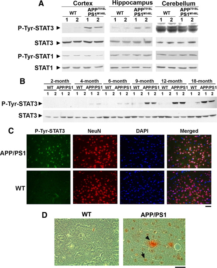
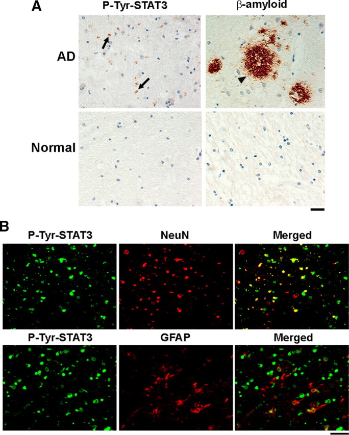
One of the pathological hallmarks of Alzheimer's disease (AD) is deposition of extracellular amyloid-beta (Abeta) peptide, which is generated from the cleavage of amyloid precursor protein (APP). Accumulation of Abeta is thought to associate with the progressive neuronal death observed in AD. However, the precise signaling mechanisms underlying the action of Abeta in AD pathophysiology are not completely understood. Here, we report the involvement of the transcription factor signal transducer and activator of transcription 3 (STAT3) in mediating Abeta-induced neuronal death. We find that tyrosine phosphorylation of STAT3 is elevated in the cortex and hippocampus of APP/PS1 transgenic mice. Treatment of cultured rat neurons with Abeta or intrahippocampal injection of mice with Abeta both induces tyrosine phosphorylation of STAT3 in neurons. Importantly, reduction of either the expression or activation of STAT3 markedly attenuates Abeta-induced neuronal apoptosis, suggesting that STAT3 activation contributes to neuronal death after Abeta exposure. We further identify Tyk2 as the tyrosine kinase that acts upstream of STAT3, as Abeta-induced activation of STAT3 and caspase-3-dependent neuronal death can be inhibited in tyk2(-/-) neurons. Finally, increased tyrosine phosphorylation of STAT3 is also observed in postmortem brains of AD patients. Our observations collectively reveal a novel role of STAT3 in Abeta-induced neuronal death and suggest the potential involvement of Tyk2/STAT3 signaling in AD pathophysiology.


Figures

Figure 1.
Figure 1.
Tyrosine phosphorylation of STAT3 is elevated in cortex and hippocampus of AD mouse models. A, Cortices, hippocampi, and cerebelli from 12-month-old APP751SL/PS1M146L double-transgenic mouse brains were lysed and subjected to Western blot analysis using p-Tyr705 STAT3 (p-Tyr-STAT3), p-Tyr701 STAT1 (p-Tyr-STAT1), and their corresponding total antibodies. B, Cortices from different ages of APPswe/PS1ΔE9 (designated as APP/PS1) transgenic mice were homogenized, and lysates were subjected to Western blot analysis for p-Tyr-STAT3 and STAT3. C, Paraffin sections of 9-month-old APP/PS1 transgenic mouse brain cortices were stained using p-Tyr-STAT3 antibody, DAPI, and the neuron-specific marker NeuN as indicated. Scale bar, 50 μm. D, Paraffin sections of 12-month-old APP/PS1 transgenic mouse cortices were stained using p-Tyr-STAT3 antibody (brown; DAB) and Aβ antibody (red; AEC). Scale bar, 50 μm. The arrow indicated the p-Tyr STAT3-positive cells, whereas the arrowhead depicted the Aβ deposit.
Figure 2.
Figure 2.
Tyrosine phosphorylation of STAT3 is increased in cortical neurons or in mouse brains in response to Aβ insult. A, Primary cortical neurons at 7 DIV were treated with Aβ25-35 (40 μm; H2O served as control) for various time periods as indicated. Cell lysates were collected and subjected to immunoblotting using p-Tyr-STAT3 and total STAT3 antibodies. B, Primary cortical neurons treated with Aβ25-35 for 1 h were fixed and immunostained using p-Tyr-STAT3 and β-tubulin-III (β-Tub III) antibodies. Scale bar, 10 μm. C, D, Tyrosine phosphorylation of STAT3 increased in mouse brains upon injection of Aβ1-42. A single intrahippocampal injection of Aβ1-42 or Aβ42-1 in mice was performed. C, Lysates of mouse cortex and hippocampus at 20 d after injection were collected and subjected to Western blot analysis for p-Tyr-STAT3 and STAT3. D, Cortex sections were collected at 20 d after injection and stained for p-Tyr-STAT3, DAPI, NeuN, or the astrocytic marker GFAP as indicated. Scale bar, 50 μm.
Figure 3.
Figure 3.
Knockdown of STAT3 ameliorates Aβ-induced neuronal apoptosis. A–D, Knockdown of STAT3 reduced neuronal cell death induced by Aβ25-35 treatment, whereas expression of RNAi-resistant STAT3 restored the neurotoxic effect of Aβ. PC12 cells were transfected with STAT3 siRNA (A, B) or with STAT3 shRNA construct alone or together with shRNA-resistant STAT3 as indicated (C, D). A, C, Western blot analysis for STAT3 was performed at 3 d after transfection of PC12 cells (α-tubulin serves as a loading control). B, D, After transfection, PC12 cells were differentiated with NGF for 4 d, and then exposed to Aβ25-35 for 24 h. The cell viability was assessed by MTT assay. B, Data are represented as mean ± SD (n = 3) (***p < 0.005, Aβ vs Con in scrambled siRNA condition; ANOVA). D, Data are represented as mean ± SEM (n ≥ 3) (*p < 0. 05, Aβ vs Con treatment; ANOVA). E–H, Blocking STAT3 activation protected the cortical neurons from Aβ-induced apoptosis. E, F, Primary cortical neurons at 7 DIV were pretreated with cell-permeable STAT3 inhibitor peptide (200 μg/ml) for 30 min before Aβ treatment. The cell viability of neurons after treatment with Aβ25-35 (E) or Aβ1-42 (F) was measured by MTT assay (*p < 0.05, Aβ vs Con in the absence of STAT3 inhibitor; ANOVA). G, H, Inhibition of STAT3 activation attenuates the increase of caspase-3 activity in Aβ25-35-treated neurons. G, The cell lysates were analyzed by immunoblotting using caspase-3 antibody. H, Quantification of the caspase-3 activity. Caspase-3 activity was determined by measuring the hydrolysis of fluorometric substrate Ac-DEVD-MCA (***p < 0.005 Aβ vs Con in the absence of STAT3 inhibitor; ##p < 0.01, STAT3 inhibitor vs control on Aβ exposure; ANOVA).
Figure 4.
Figure 4.
Inhibition of STAT3 activation reduces Aβ-induced gene transcription. A, Aβ25-35 treatment increased the transcriptional activity of STAT3 in PC12 cells. PC12 cells were transiently transfected with a STAT3 (pSTAT3-TA-Luc) or STAT1 (pGAS-TA-Luc) reporter gene construct and an internal control plasmid (β-gal-pCMV). Luciferase activity was measured and normalized against the β-gal activity in the samples. Promoter activity was expressed as the ratio of luciferase activity. Data are represented as mean ± SD (n = 4) (***p < 0.005, Aβ or NGF treatment vs Con in pSTAT3-transfected condition; ###p < 0.005, NGF vs Con in pGAS-transfected condition; ANOVA). B, Cortical neurons were pretreated with STAT3 inhibitor peptide for 30 min, followed by Aβ25-35 (40 μm) treatment for 6 h. Total RNA was collected and reverse transcribed. The cDNA was subjected to real-time PCR analysis using iNOS-specific primers. Data are represented as mean ± SEM; n = 3. Results were analyzed with ANOVA (**p < 0.01, Aβ vs Con; #p < 0.05, STAT3 inhibitor vs control upon Aβ exposure). C, iNOS and TRAIL mRNA expression increased in APP/PS1 mouse forebrains. Forebrains of APP/PS1 mice at 6 months were collected and the mRNA expression of iNOS or TRAIL from wild-type and APP/PS1 mice was determined by quantitative PCR. Data are represented as mean ± SEM; n = 3. Results were analyzed with Student's t test (***p < 0.005).
Figure 5.
Figure 5.
Tyk2 is required for the Aβ-induced tyrosine phosphorylation of STAT3 and neuronal cell death. A, Primary cortical neurons were pretreated for 2 h with various inhibitors including a potent inhibitor of JAK (JAK inhibitor 1), a JAK2 inhibitor (AG490), a Src tyrosine kinase inhibitor (PP2), and a protein synthesis inhibitor [cycloheximide (Cyclohex)], followed by treatment with Aβ25-35 for 4 or 24 h. The cell lysates were immunoblotted with p-Tyr-STAT3 and total STAT3 antibodies. B, Primary cortical neurons were treated with Aβ25-35. The cell lysates were coimmunoprecipitated using Tyk2 or JAK2 antibodies, and then subjected to Western blot analysis using phosphotyrosine antibody (p-Tyr). C–F, Primary cortical neurons prepared from tyk2−/− mice were treated with Aβ25-35. C, The cell lysates were subjected to immunoblotting using antibodies against p-Tyr-STAT3, total STAT3, caspase-3, or α-tubulin as indicated. Quantification analysis of fold change in p-Tyr-STAT3 (D) and cleaved caspase-3 (E) (*p < 0.05, ***p < 0.005, Aβ treatment for various time period vs 0 h in WT neurons; #p < 0.05, ###p < 0.005, tyk2−/− neurons vs WT neurons at specific time periods; ANOVA). F, Cell viability of tyk2−/− neurons after treatment with Aβ was measured by MTT assay (###p < 0.001, tyk2−/− vs WT neurons on Aβ treatment; ANOVA). Error bars indicate SEM.
Figure 6.
Figure 6.
Tyrosine phosphorylation of STAT3 is increased in the hippocampus of Alzheimer's disease patients. A, Paraffin sections of postmortem brains from AD patients were immunostained with Aβ and p-Tyr-STAT3 antibodies. Cell nuclei were stained with hematoxylin. The arrows indicated the p-Tyr STAT3-positive cells, and the arrowhead indicated the β-amyloid deposit (n = 5 for the normal and n = 7 for the AD patient group). Scale bar, 50 μm. B, Paraffin sections of postmortem brains from AD patients were costained with p-Tyr-STAT3 antibody and NeuN or GFAP antibodies. Scale bar: left panels, 50 μm.

References

    1. Akama KT, Albanese C, Pestell RG, Van Eldik LJ. Amyloid β-peptide stimulates nitric oxide production in astrocytes through an NFκB-dependent mechanism. Proc Natl Acad Sci U S A. 1998;95:5795–5800. - PMC - PubMed
    1. Akira S. Roles of STAT3 defined by tissue-specific gene targeting. Oncogene. 2000;19:2607–2611. - PubMed
    1. Arendash GW, King DL, Gordon MN, Morgan D, Hatcher JM, Hope CE, Diamond DM. Progressive, age-related behavioral impairments in transgenic mice carrying both mutant amyloid precursor protein and presenilin-1 transgenes. Brain Res. 2001;891:42–53. - PubMed
    1. Borchelt DR, Davis J, Fischer M, Lee MK, Slunt HH, Ratovitsky T, Regard J, Copeland NG, Jenkins NA, Sisodia SS, Price DL. A vector for expressing foreign genes in the brains and hearts of transgenic mice. Genet Anal. 1996;13:159–163. - PubMed
    1. Buxbaum JD, Gandy SE, Cicchetti P, Ehrlich ME, Czernik AJ, Fracasso RP, Ramabhadran TV, Unterbeck AJ, Greengard P. Processing of Alzheimer β/A4 amyloid precursor protein: modulation by agents that regulate protein phosphorylation. Proc Natl Acad Sci U S A. 1990;87:6003–6006. - PMC - PubMed

Publication types

MeSH terms