. 2010 May 19;30(20):6930-43.
doi: 10.1523/JNEUROSCI.5395-09.2010.

Rac1 regulates neuronal polarization through the WAVE complex

Affiliations

Rac1 regulates neuronal polarization through the WAVE complex

Sabina Tahirovic et al. J Neurosci. .

Abstract

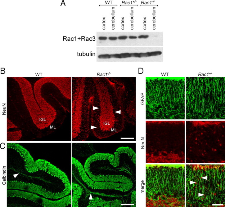
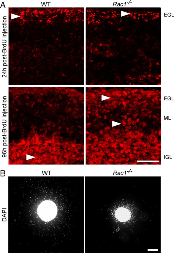
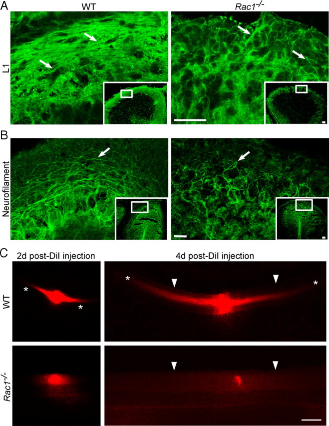
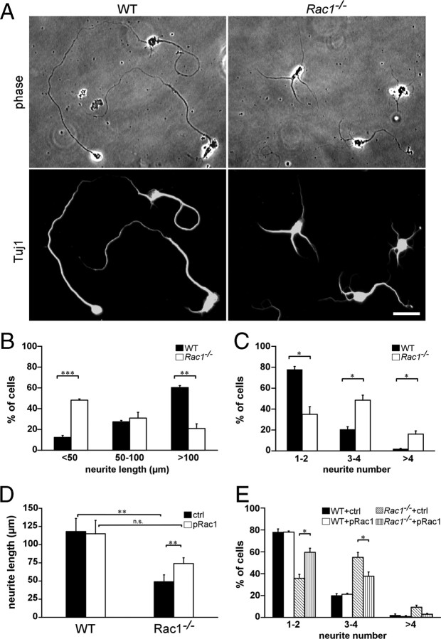
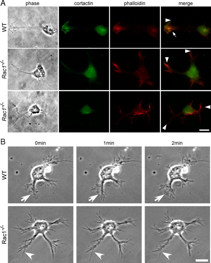
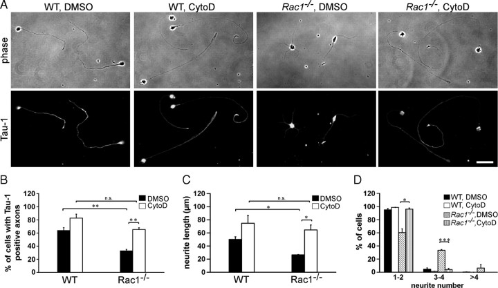
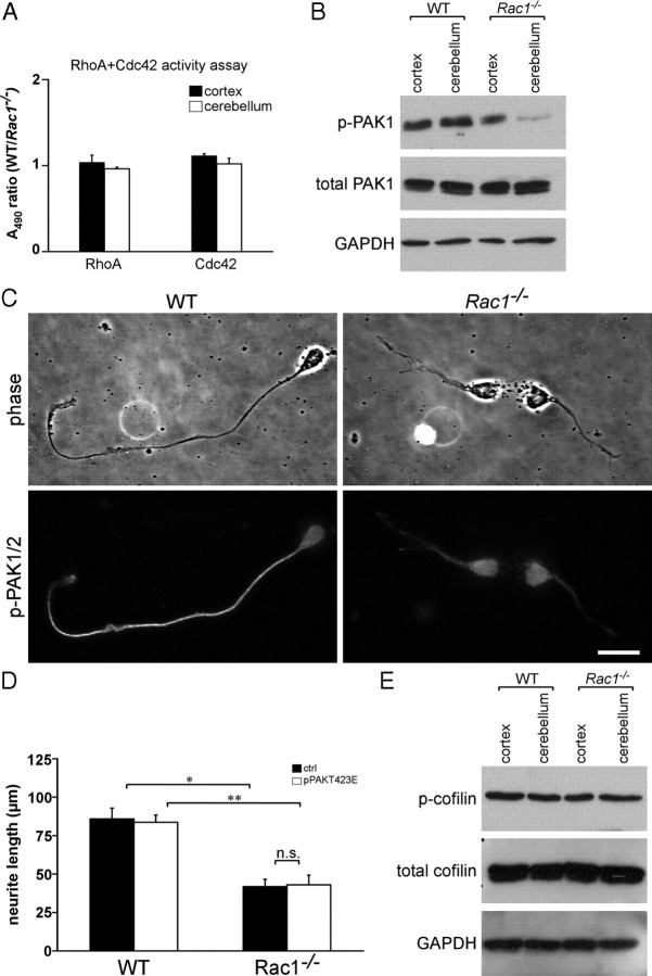
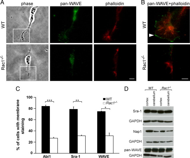
Neuronal migration and axon growth, key events during neuronal development, require distinct changes in the cytoskeleton. Although many molecular regulators of polarity have been identified and characterized, relatively little is known about their physiological role in this process. To study the physiological function of Rac1 in neuronal development, we have generated a conditional knock-out mouse, in which Rac1 is ablated in the whole brain. Rac1-deficient cerebellar granule neurons, which do not express other Rac isoforms, showed impaired neuronal migration and axon formation both in vivo and in vitro. In addition, Rac1 ablation disrupts lamellipodia formation in growth cones. The analysis of Rac1 effectors revealed the absence of the Wiskott-Aldrich syndrome protein (WASP) family verprolin-homologous protein (WAVE) complex from the plasma membrane of knock-out growth cones. Loss of WAVE function inhibited axon growth, whereas overexpression of a membrane-tethered WAVE mutant partially rescued axon growth in Rac1-knock-out neurons. In addition, pharmacological inhibition of the WAVE complex effector Arp2/3 also reduced axon growth. We propose that Rac1 recruits the WAVE complex to the plasma membrane to enable actin remodeling necessary for axon growth.


Figures

Figure 1.
Figure 1.
Loss of Rac1 from the nervous system results in defects in the migration of CGNs. A, Depletion of Rac1 from Rac1−/− cerebellum. P9 cerebellar and cortical protein extracts were prepared from wild-type, Rac1+/−, and Rac1−/− brains. Equal amounts of protein were subjected to Western blot analysis using an antibody recognizing Rac1 and cross-reacting with Rac3. The signal detected in cortical extracts of Rac1−/− brains represents Rac3, which is absent from cerebellar extracts. B, Ablation of Rac1 causes aberrant distribution of NeuN-positive neurons. Sagittal sections from P18 cerebella were immunostained with NeuN. Many CGNs stacked in the ML of the Rac1-null cerebellum can be observed (arrowheads). In contrast, in the wild type, the majority of CGNs have reached the IGL. Scale bar, 150 μm. C, Sagittal sections from P18 cerebella immunostained with calbindin (axonal tracts of Purkinje neurons, arrowheads). The Rac1-null cerebellum shows the typical monolayer of Purkinje neurons. Scale bar, 200 μm. D, Sagittal sections from P18 cerebella coimmunostained with NeuN and GFAP. The Rac1−/− cerebellum exhibits a normal organization of GFAP-positive Bergmann glia cells that are in proximity to NeuN-positive CGNs (arrowheads). Scale bar, 40 μm.
Figure 2.
Figure 2.
Abnormal in vivo and in vitro migration of Rac1−/− CGNs. A, Dividing cerebellar granule cell precursors were labeled by systemic injection of BrdU into P7 wild-type and Rac1-deficient mice. At 24 h after BrdU injection, the majority of BrdU-labeled granule cells are in the EGL in both the wild-type and Rac1−/− cerebellum (arrowheads). At 96 h after injection, the majority of BrdU-labeled granule cells in the wild-type cerebellum have reached the IGL (arrowhead). By contrast, in the Rac1−/− cerebellum, more BrdU-labeled granule cells (arrowheads) stayed in the EGL and ML. Scale bar, 50 μm. B, EGL explants from P5 wild-type and Rac1−/− mice were stained with DAPI after 2 DIV. Rac1−/− neurons migrate from the explant less efficiently than wild-type neurons. Scale bar, 200 μm.
Figure 3.
Figure 3.
Rac1−/− neurons show deficient axon extension in vivo. A, P15 coronal sections from the middle part of the cerebellum immunostained with an antibody against L1 that visualizes parallel fibers in the ML. Images of boxed regions of the ML are depicted at higher magnification. L1 immunostaining reveals less densely packed parallel fibers in the ML of the Rac1-null cerebellum compared with wild type (arrows). Scale bars, 30 μm. B, P18 midsagittal sections immunostained with an antibody against neurofilament to visualize axons. Images of boxed regions of the ML are depicted at higher magnification. Long axonal fibers can be observed in the wild-type cerebellum and short and misrouted axons in the Rac1−/− cerebellum (arrows). Scale bars, 30 μm. C, DiI-labeled parallel fibers in P15 coronal sections of wild-type and Rac1−/− cerebella. At 2 d after DiI injection, parallel fiber labeling within the ML is visible in the wild-type cerebellum (asterisks), but not in the Rac1−/− cerebellum. At 4 d after injection, long parallel fibers project from both sides of the DiI injection place in the wild-type cerebellum, but this labeling is absent in the Rac1−/− cerebellum. The pial surface of the cerebellum is indicated by arrowheads. Note that the intensity of DiI labeling at the injection site in wild-type and Rac1−/− cerebellum is comparable (2 d after DiI injection). Scale bar, 100 μm.
Figure 4.
Figure 4.
Reduced length and an increased number of neurites in cultured Rac1−/− CGNs can be reversed by overexpressing Rac1. A, Dissociated CGNs were immunostained after 2 DIV with an antibody against Tuj1. Rac1−/− CGNs display several shorter Tuj1-positive neurites, whereas wild-type CGNs typically exhibit one or two longer Tuj1-positive neurites. Scale bar, 25 μm. B, Quantification of the length of the longest Tuj1-stained neurite. The values represent averages ± SEM from three independent cultures (n = 100 cells/culture; ***p < 0.001; **p < 0.01). C, Quantification of the neurite number. The values represent averages ± SEM from three independent cultures (n = 100 cells/culture; *p < 0.05). D, Quantification of the length of the longest neurite in CGNs cotransfected with pEYFP-C1 and pcDNA3.1 (ctrl) or with pEYFP-C1 and pRac1 (pRac1) at 3 DIV. The values represent averages ± SEM from four independent cultures (n = 100 cells/culture; **p < 0.01). E, Quantification of the neurite number in CGNs cotransfected with pEYFP-C1 and pcDNA3.1 (ctrl) or with pEYFP-C1 and pRac1 (pRac1) at 3 DIV. The values represent averages ± SEM from three independent cultures (n = 100 cells/culture; *p < 0.05).
Figure 5.
Figure 5.
Rac1−/− CGNs lack lamellipodia and show reduced actin dynamics. A, Dissociated CGNs at 1.5 DIV were costained with rhodamine–phalloidin to visualize actin filaments and with an antibody against cortactin to visualize lamellipodia. Images from wild-type and two different Rac1−/− neurons are shown. Cortactin and phalloidin staining of a wild-type neuron reveal a lamellipodium (arrow) and a filopodium (arrowhead), whereas Rac1−/− neurons only have filopodia at their neurite tips (arrowheads) but lack lamellipodia. Scale bar, 10 μm. B, Time-lapse videomicroscopy of representative wild-type and Rac1−/− CGNs at 1.5 DIV. Three frames at 1 min intervals are shown. A dynamic lamellipodium of a wild-type growth cone is indicated by an arrow. A filopodium of a Rac1−/− growth cone is indicated by an arrowhead. Scale bar, 10 μm.
Figure 6.
Figure 6.
Actin depolymerization reverses Rac1-mediated defects in axon growth. A, Dissociated CGNs were treated with cytochalasin D (CytoD) or control treated with DMSO 4 h after plating. Cells were fixed at 2 DIV and immunostained for the axonal marker Tau-1. CytoD-treated Rac1−/− neurons extend longer, Tau-1-positive axons bearing a typical proximodistal staining gradient. DMSO-treated Rac1−/− neurons extend several shorter, Tau-1-negative neurites. Cytochalasin D treatment abrogates the difference in axon formation between Rac1−/− and wild-type CGNs. Scale bar, 30 μm. B, Quantification of axon formation. Cells, bearing at least one Tau-1-positive axon (proximodistal staining gradient), were scored as positive. The values represent averages ± SEM from three independent cultures (n = 100 cells/culture; **p < 0.01). C, D, Dissociated CGNs were treated with CytoD or DMSO 4 h after plating. Cells were fixed at 1 DIV and visualized by immunostaining for Tuj1. Quantification of the length of the longest Tuj1-stained neurite (C) or neurite number (D). The values represent averages ± SEM from three independent cultures (n = 100 cells/culture; *p < 0.05; ***p < 0.001).
Figure 7.
Figure 7.
Rac1 deletion results in a reduced activity of PAK but does not change the activity of cofilin. A, ELISA-based assay for RhoA and Cdc42 activation. The results are expressed as ratios between A490 in wild-type and Rac1−/− extracts. The values represent averages ± SEM from three animals. There is no significant difference between the activity of RhoA or Cdc42 in Rac1−/− and wild-type cerebella. Cortical extracts were used as control. B, Analysis of phospho-PAK1 (p-PAK1) and PAK1 protein levels from P9 cortices and cerebella. Rac1−/− cerebellar extracts show reduced levels of p-PAK1 compared with wild-type extracts. C, Dissociated CGNs at 2 DIV were immunostained with an antibody against p-PAK1/2. A decreased level of p-PAK1/2 can be observed in neurites of Rac1−/− CGNs. By contrast, higher levels of p-PAK1/2 accumulate in a neurite of a wild-type CGN. Scale bar, 15 μm. D, Quantification of the length of the longest neurite in CGNs cotransfected with pEYFP-C1 and pcDNA3.1/myc-His A (ctrl) or pEYFP-C1 and pPAKT423E (pPAKT423E) at 3 DIV. The values represent averages ± SEM from three independent cultures (n = 100 cells/culture; *p < 0.05; **p < 0.01). Overexpression of constitutively active PAK1 (PAKT423E) in Rac1−/− CGNs does not increase neurite length. E, Analysis of phosphorylated-cofilin (p-cofilin) and cofilin protein levels from P9 cortices and cerebella. Wild-type and Rac1−/− extracts show comparable levels of p-cofilin.
Figure 8.
Figure 8.
Rac1−/− neurons mislocalize WAVE from the growth cone plasma membrane. A, Dissociated CGNs at 1 DIV were costained with rhodamine–phalloidin and an antibody against WAVE (pan-WAVE). Rac1−/− neurons that still contained some lamellipodia-like protrusions were examined for WAVE localization. Scale bar, 10 μm. B, Merged images of boxed regions in A are depicted at higher magnification. WAVE is absent from the edges of Rac1−/− growth cones, whereas in wild-type neurons WAVE predominantly localizes to membranes of lamellipodial protrusions (arrowhead). Scale bar, 5 μm. C, Quantification of plasma membrane localization of Abi1, Sra-1, and WAVE. CGNs, showing plasma membrane localization of the respective protein in at least one growth cone, were scored as positive. The values represent averages ± SEM from three independent cultures (n = 100 cells/culture; *p < 0.05; **p < 0.01; ***p < 0.001). D, Mislocalization of the WAVE complex in the Rac1−/− neurons is not caused by protein degradation. Western blot analysis of P9 cortical and cerebellar extracts reveals no difference in the protein amounts of the respective WAVE complex components.
Figure 9.
Figure 9.
WAVE complex and its downstream effector Arp2/3 regulate axon growth downstream of Rac1. A, Dissociated CGNs were cotransfected with pEYFP-C1 and pcDNA3.1/myc-His A (ctrl) or with pEYFP-C1 and pWAVE-CAAX (pWAVE-CAAX). Cells were fixed at 3 DIV and immunostained for Tau-1 to visualize axons. On overexpression of WAVE-CAAX, Rac1−/− CGNs extend significantly more Tau-1-positive axons bearing a typical proximodistal staining gradient. Instead, Rac1−/− neurons transfected with the ctrl typically extend several shorter, Tau-1-negative neurites. Overexpression of WAVE-CAAX partially rescues the axon formation defect of Rac1−/− CGNs. Scale bar, 25 μm. B, Quantification of axon formation on transfection of pWAVE-CAAX or ctrl. Cells, bearing at least one Tau-1-positive axon (proximodistal staining gradient), were scored as positive. The values represent averages ± SEM from four independent cultures (n = 100 cells/culture; **p < 0.01; ***p < 0.001). C, Quantification of the length of the longest neurite in CGNs transfected with pWAVE-CAAX or ctrl. The values represent averages ± SEM from five independent cultures (n = 100 cells/culture; **p < 0.01). D, Time-lapse videomicroscopy of representative growth cones from wild-type CGNs cultured for 1 d and imaged before and after the addition of the Arp2/3 inhibitor (CK-548). The top panel represents four frames at 30 s intervals before the addition of CK-548, and the bottom panel, after the addition of CK-548. The additional two frames depict the growth cone dynamics 6 min before and after the inhibitor was added. Scale bar, 10 μm.

Similar articles

Cited by

References

    1. Albertinazzi C, Gilardelli D, Paris S, Longhi R, de Curtis I. Overexpression of a neural-specific rho family GTPase, cRac1B, selectively induces enhanced neuritogenesis and neurite branching in primary neurons. J Cell Biol. 1998;142:815–825. - PMC - PubMed
    1. Benesch S, Lommel S, Steffen A, Stradal TE, Scaplehorn N, Way M, Wehland J, Rottner K. Phosphatidylinositol 4,5-biphosphate (PIP2)-induced vesicle movement depends on N-WASP and involves Nck, WIP, and Grb2. J Biol Chem. 2002;277:37771–37776. - PubMed
    1. Bogdan S, Grewe O, Strunk M, Mertens A, Klämbt C. Sra-1 interacts with Kette and Wasp and is required for neuronal and bristle development in Drosophila. Development. 2004;131:3981–3989. - PubMed
    1. Bokoch GM. Regulation of innate immunity by Rho GTPases. Trends Cell Biol. 2005;15:163–171. - PubMed
    1. Bolis A, Corbetta S, Cioce A, de Curtis I. Differential distribution of Rac1 and Rac3 GTPases in the developing mouse brain: implications for a role of Rac3 in Purkinje cell differentiation. Eur J Neurosci. 2003;18:2417–2424. - PubMed

Publication types

MeSH terms