. 2010 Jun 8;107(23):10596-601.
doi: 10.1073/pnas.0913280107. Epub 2010 May 19.

Conversion of a yeast prion protein to an infectious form in bacteria

Affiliations

Conversion of a yeast prion protein to an infectious form in bacteria

Sean J Garrity et al. Proc Natl Acad Sci U S A. .

Abstract

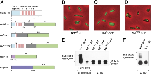
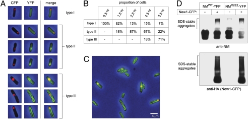
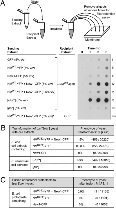
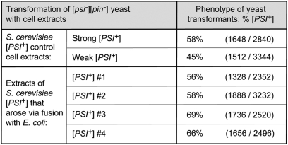
Prions are infectious, self-propagating protein aggregates that have been identified in evolutionarily divergent members of the eukaryotic domain of life. Nevertheless, it is not yet known whether prokaryotes can support the formation of prion aggregates. Here we demonstrate that the yeast prion protein Sup35 can access an infectious conformation in Escherichia coli cells and that formation of this material is greatly stimulated by the presence of a transplanted [PSI(+)] inducibility factor, a distinct prion that is required for Sup35 to undergo spontaneous conversion to the prion form in yeast. Our results establish that the bacterial cytoplasm can support the formation of infectious prion aggregates, providing a heterologous system in which to study prion biology.


Conflict of interest statement

The authors declare no conflict of interest.

Figures

Fig. 1.
Fig. 1.
Behavior of yeast prion-forming domain fusion proteins in E. coli cells. (A) Fusion constructs. Indicated residues from yeast prion-forming proteins, Sup35 (NM), New1, or Rnq1, were fused to a fluorescent protein (FP). FPs used are GFP, YFP, and CFP. The prion-forming domain of NM is enlarged to highlight its Q/N-rich segment and five complete copies of an oligopeptide repeat sequence; NM-FP lacks repeats 2–5, and NMR2E2-FP has two additional copies of repeat 2. Constructs containing CFP have three HA-epitope tags fused at the C-terminal end. Fusion genes encoding either the GFP or YFP moiety were expressed under the control of the same arabinose-inducible promoter, whereas genes encoding the CFP moiety were expressed under the control of an isopropyl beta-D-thiogalactopyranoside (IPTG)-inducible promoter. (B–D) Fluorescence images of cells containing the indicated NM-GFP fusion protein. The cells were transformed with plasmids encoding each fusion protein under the control of an inducible promoter. The images show cells examined after the induction of fusion protein synthesis for 5 h. Foci were not observed in cells containing the NM-GFP fusion protein. Images are representative fields; in each case, several hundred cells were examined. (E) SDD-AGE analysis of cell extracts containing the indicated NM-GFP fusion protein. Blot was probed with an anti-GFP antibody. SDS-stable aggregates migrate as smears above soluble material. Soluble NM-GFP fusion protein (from S. cerevisiae as well as E. coli) migrates as multiple species. Extracts were examined by SDD-AGE analysis in at least three experiments, with similar results. (F) SDD-AGE analysis of cell extracts containing the indicated fusion protein. Blot was probed with an anti-GFP antibody. SDS-stable aggregates migrate as smears above the soluble material. SDS/PAGE and Western blot analysis of cell extracts containing these fusion proteins as well as the NM-GFP fusion protein (A) suggests that observed differences in aggregation behavior are unlikely due to differences in intracellular fusion protein levels (Fig. S1B). Extracts were examined by SDD-AGE analysis in at least three experiments, with similar results.
Fig. 2.
Fig. 2.
Behavior of NM-YFP fusion protein in E. coli cells also containing New1-CFP fusion protein. Cells were transformed with two compatible plasmids, one encoding the NMWT-YFP fusion protein under the control of an arabinose-inducible promoter and the other encoding the New1-CFP fusion protein under the control of a leaky IPTG-inducible promoter. The cells were grown in the absence of IPTG and arabinose was added to induce the synthesis of the NMWT-YFP fusion protein. Sufficient New1-CFP fusion protein was produced under these conditions to form detectable fluorescent foci and SDS-stable material. (A) Fluorescence images of representative cells containing both fusion proteins, 1.5 h after induction of NM-YFP fusion protein synthesis (type I), 2.5 h after induction of NM-YFP fusion protein synthesis (type II), and 5.5 h after induction of NM-YFP fusion protein synthesis (type III). Shown are CFP channel images, YFP channel images, and merged images. (B) Table showing proportion of cells of each type (I, II, or III, as shown in A) at indicated times after induction of NM-YFP fusion protein synthesis. (C) Fluorescence image of a field of cells containing both fusion proteins, 5.5 h after induction of NM-YFP fusion protein synthesis. Similar ribbon-like structures were observed in cells containing New1-CFP together with NMR2E2-YFP (Fig. S3) (D) SDD-AGE analysis of cell extracts containing either NM-YFP or both NM-YFP and New1-CFP, 5.5 h after induction of NM-YFP fusion protein synthesis. SDS-stable NM-YFP aggregates migrate near the top of the gel. Blot was probed with anti-NM antibody to detect NM-YFP (Upper) and anti-HA antibody to detect New1-CFP (which bears three copies of an HA tag at its C terminus) (Lower). Extracts were examined by SDD-AGE in at least two experiments, with similar results.
Fig. 3.
Fig. 3.
Cells with both NM-YFP and New1-CFP aggregates contain seeding-competent, infectious material. (A) E. coli cell extracts containing SDS-soluble NM-GFP were seeded with E. coli cell extract containing overproduced GFP, overproduced NM-YFP, overproduced New1-CFP, or overproduced NM-YFP and New1-CFP; yeast cell extract prepared from either a [PSI+] or a [psi] strain was also used as seed (strains YJW96 and SG775, respectively). A seed-only control sample (*) consisted of E. coli cell extract containing overproduced NM-YFP and New1-CFP diluted into E. coli extract containing overproduced GFP only. Cartoon depicts experimental protocol. Samples from seeded reactions were removed at various time points, treated with 2% SDS, and filtered through a cellulose acetate (low-binding) membrane. SDS-stable aggregates that were retained were probed with anti-NM antibody. Extracts were examined for seeding activity in three experiments, with similar results. (B) Infection of [pin][psi] yeast spheroplasts with extract prepared from E. coli cells containing the indicated fusion proteins, or with extract prepared from either [PSI+] or [psi] yeast cells (strains YJW96 and SG775, respectively). The New1-CFP fusion protein was encoded on the chromosome under the control of an IPTG-inducible promoter, and NMR2E2-YFP fusion protein was encoded on a plasmid under the control of an arabinose-inducible promoter. Extracts were prepared from cells 6.5 h after induction of fusion protein synthesis with IPTG and arabinose. Yeast spheroplasts were cotransformed with a yeast shuttle vector containing a URA+ selectable marker. [PSI+] transformants exhibited primarily a “strong” phenotype (3). (C) Fusion of [pin][psi] yeast spheroplasts with protoplasts prepared from E. coli cells containing the indicated fusion proteins. The New1-CFP fusion protein was encoded on the chromosome under the control of an IPTG-inducible promoter; the NMR2E2-YFP fusion protein was encoded on a plasmid under the control of an arabinose-inducible promoter. Protoplasts were prepared 6.5 h after induction of fusion protein synthesis with IPTG and arabinose. E. coli cells also contained a yeast shuttle vector with a URA+ selectable marker. Analysis of these data by Fisher's exact test suggests that the observed difference in the frequencies of [PSI+] transformants is statistically significant (P = 10−5). All [PSI+] transformants exhibited a “strong” phenotype (3) (Fig. S4).
Fig. 4.
Fig. 4.
Material derived from [PSI+] yeast strains that arose via fusion with E. coli protoplasts is similarly infectious to material derived from control [PSI+] yeast strains. Extracts of four representative [PSI+] yeast strains obtained via fusion with E. coli protoplasts (labeled [PSI+] #1–4) as well as control extracts from both strong (SG862) and weak (SG863) [PSI+] yeast strains were used to transform [pin] [psi] yeast cells. Frequency of [PSI+] observed (as percentage of total transformants) is shown. Strain details are given in Table S1; colony phenotypes are shown in Fig. S4.

Similar articles

Cited by

References

    1. Aguzzi A, Polymenidou M. Mammalian prion biology: One century of evolving concepts. Cell. 2004;116:313–327. - PubMed
    1. Wickner RB, Liebman SW, Saupe SJ. Prions of yeast and filamentous fungi: [URE3+], [PSI+], [PIN+], and [Het-s] In: Prusiner SB, editor. Prion Biology and Diseases. Cold Spring Harbor Lab Press: Cold Spring Harbor, NY; 2004. pp. 305–372.
    1. Chien P, Weissman JS, DePace AH. Emerging principles of conformation-based prion inheritance. Annu Rev Biochem. 2004;73:617–656. - PubMed
    1. Pan KM, et al. Conversion of alpha-helices into beta-sheets features in the formation of the scrapie prion proteins. Proc Natl Acad Sci USA. 1993;90:10962–10966. - PMC - PubMed
    1. Kimura Y, Koitabashi S, Fujita T. Analysis of yeast prion aggregates with amyloid-staining compound in vivo. Cell Struct Funct. 2003;28:187–193. - PubMed

Publication types

MeSH terms

LinkOut - more resources